[ad_1]
Ex vivo phage show towards XFS supplies
Peptide sequences that bind to XFS supplies had been deduced from the DNA sequences of chosen phage clones. Three rounds of biopanning towards XFS supplies sourced from completely different sufferers yielded an enrichment of two peptides: LPSYNLHPHVPP (p-LPS) and IPLLNPGSMQLS (p-IPL) (Further file 1: Desk S1). The excessive diploma of selectivity of those phages in direction of XFS supplies was aided by: i. unfavourable screening by removing of phage that certain regular lenses by unfavourable biopanning, and ii. removing of XFS supplies from the lens capsules, leading to particular amplification of phage that solely certain to those fibrils. Furthermore, the strong nature of the binding was evident as samples got here from a number of various sufferers but the responses had been comparable. Lastly, using extracted human aqueous humor at 37 °C [26] was important to those experiments in order to keep up an answer atmosphere (i.e., pH, ionic energy, proteins, osmolarity) that was as shut as attainable to the physiological atmosphere the place the focusing on of fibrils would happen.
Evaluating the focusing on means of phage-displayed peptides
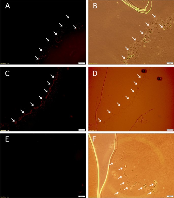
Human lens capsules with XFS supplies had been stained with fluorescently labeled wild-type phages in addition to phages displaying the enriched peptides. It is very important word that the conjugated fluorophore (Cy5 NHS ester dye) molecule didn’t intervene with phage binding to the XFS supplies because it reacts with the first amino group of lysine, which was not current within the enriched peptides. Each phage-displayed peptides (p-LPS and p-IPL) confirmed particular binding to XFS supplies (Fig. 1A, C), the place the presence of XFS supplies was already confirmed utilizing bright-field microscopy (Fig. 1B, D, F). Wild-type phages confirmed no noticeable interplay with XFS supplies on the floor of the lens capsule (Fig. 1E). XFS supplies don’t cowl the entire floor of the lens cuspule, which means that labeled phages had the prospect to work together with the non-XFS altered areas of the lens capsule. Phage had been noticed to bind solely to the XFS areas of the lens capsule, additional confirming their specificity in direction of the XFS supplies.
Localization of labeled phage-displayed peptides on the human lens capsule having XFS supplies. M13 phages with XFS material-targeting peptides displayed on coat protein pIII and wild-type M13 phages had been labeled with Cy5 fluorescent dye and incubated with human lens capsule containing XFS supplies. Phages with displayed p-LPS (A) and p-IPL (C) peptides on their floor and labeled with Cy5 dye had been selectively certain to the XFS supplies on the lens capsule. Wild-type phages labeled with Cy5 dye (management) didn’t present any particular interplay with XFS supplies (E). The presence of XFS supplies on the lens capsule was confirmed within the vibrant discipline mode of the microscope (B, D, and F). Arrows present the exfoliative zones on the human lens capsule. (Scale bars = 100 µm)
Peptide-particle conjugation
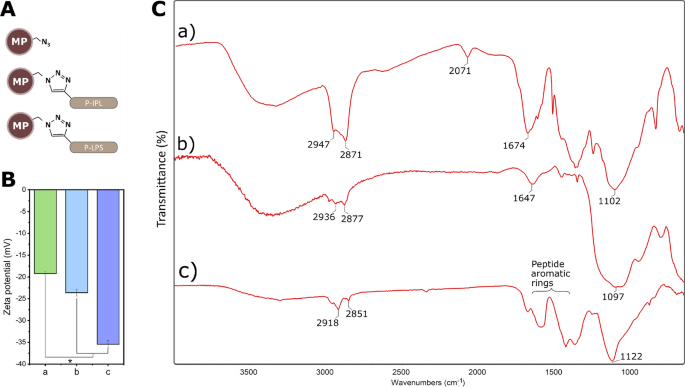
Conjugation of alkyne-modified peptides to azide-functionalized MPs (Fig. 2A) was confirmed with floor zeta potential measurements, Fourier-transform infrared spectroscopy (FTIR), and a aggressive inhibition assay. Peptide tethering by the N-terminal area was anticipated to yield a rise in unfavourable cost on the floor of the MPs, as was noticed (Fig. 2B). FTIR spectrum from MPs earlier than conjugation to peptides confirmed a transmittance peak round 2071 cm−1, which is attributed to the uneven stretching vibration of the free azide teams (Fig. 2C. a). The free azide group was absent within the spectra of peptide-conjugated MP samples as a result of conversion of free azide teams to triazole ring throughout azide-alkyne cycloaddition (Fig. 2C. b, c). The height noticed at 1647 cm−1 in p-IPL might symbolize carbonyl teams of amide bonds of the peptide (Fig. 2C. b). The bands at wavelengths between 1400–1650 cm−1 correspond to fragrant rings discovered on p-LPS. (Fig. 2C. c).
Conjugation of peptides to MPs. (A) Schematic illustration of MPs with and with out peptide conjugates. (B) Floor zeta potential measurements and (C) FTIR spectra of MPs earlier than and after peptide conjugation to MPs. a) Azide-functionalized MPs with out peptide conjugates, b) MP-p-IPL, c) MP-p-LPS. (* represents p < 0.05, information symbolize imply ± 1 SD, n ≥ 3)
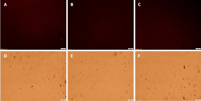
Conjugation of peptides to MPs was confirmed by labeling particles with or with out conjugated peptides with an azide-reactive dye (TAMRA alkyne fluorophore), the place subsequent fluorescence indicated that unreacted azides had been current on the MP floor (Fig. 3). MPs had been reacted with p-IPL (Fig. 3B) or p-LPS peptides (Fig. 3C), these MPs had no fluorescence in comparison with the management MPs that had no conjugated peptides (Fig. 3). Given the extreme quantity of peptides used to react with out there azides on the magnetic particles, coupled with the shortage of any unreacted azides (Fig. 3B, C), it’s affordable to conclude that just about 100% of all azides reacted to covalently tether peptides to the magnetic particles.’
Aggressive labeling of MPs with TAMRA dye. Peptide-conjugated MPs and azide-functionalized MPs with out conjugated peptides had been labeled with TAMRA dye. (A, D) Management particle clumps having free azide teams confirmed noticeably increased fluorescence below the microscope. MPs conjugated to p-IPL (B, E) and p-LPS (C, F) confirmed no fluorescence below the microscope. High pictures had been taken within the fluorescence mode utilizing TRITC filter and backside pictures had been taken within the vibrant discipline mode to verify the presence of particles and clumps of particles. (Scale bars = 50 µm)
Concentrating on functionality of MP-peptide conjugates
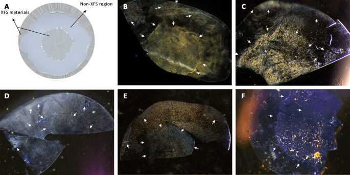
It is very important word that though there could be patient-to-patient variations within the XFS deposition sample on the lens capsule, typically it’s distributed with a non-XFS intermediate zone that separates exfoliative central and peripheral zones (Fig. 4A) [11]. Particular focusing on of XFS supplies was evaluated by incubating MP-p-IPL, MP-p-LPS, MP-scrambled peptides (management), or unmodified MP (management) with XFS affected lens capsules in equal quantity of extracted aqueous humor fluid and BSS irrigating answer at 37 °C. Particular binding of MP-p-IPL and MP-p-LPS to XFS supplies was noticed (Fig. 4B, C). This confirms that MP-peptide conjugates resulted in comparable binding patterns as that noticed for simply fluorescently labeled phage-displayed peptides (Fig. 1). Whereas, scrambled peptide complexes confirmed a non-specific binding and the entire floor of the lens capsule having was coated with MPs no matter XFS presence (Fig. 4D, E). The opposite management experiment utilizing virgin MPs resulted in massive clumps of MPs on the central zone (Fig. 4F).
Concentrating on of XFS supplies on the floor of the human lens capsule with MP-peptide conjugates. (A) Illustration of the anterior lens capsule exhibiting common sample of XFS deposits on its floor. (B) MP-p-IPL confirmed particular binding to XFS deposits on each central and the peripheral zones of tissue having XFS supplies. The non-XFS space of lens capsule (clean spots) confirmed much less or no particles in comparison with the exfoliated space. (C) MP-p-LPS additionally confirmed particular focusing on of XFS supplies on the floor of the lens capsule. (D) Scrambled MP-p-IPL interacted non-specifically with each XFS deposits and the world of lens capsule floor with out XFS deposits. (E) Scrambled MP-p-LPS additionally confirmed non-specific interplay with XFS supplies. (F) MP with out peptide conjugates. As proven within the picture, there’s a excessive quantity of XFS supplies on the central a part of the human lens capsule, nevertheless, there’s not sufficient binding of MPs to the XFS space. Arrows point out approximate areas of XFS supplies in every tissue pattern
Impact of magnetic discipline on XFS supplies
Micron-sized, iron oxide particles had been used as they’re the one steel oxide particle clinically authorized for biomedical purposes, much less vulnerable to nonspecific mobile uptake or vascular egress, quickly cleared (< 5 min) by the liver and spleen, and have a excessive labeling valency that enhances their binding affinity to molecular targets [21, 27,28,29,30]. These particles are biodegradable, the place particles (> 150 nm) are captured by phagocytic cells and their coating cleaved by lysosomal enzymes, and the iron oxide core is degraded into iron and oxygen by mechanisms concerned in iron metabolism [31, 32].
Magnetic pin take a look at
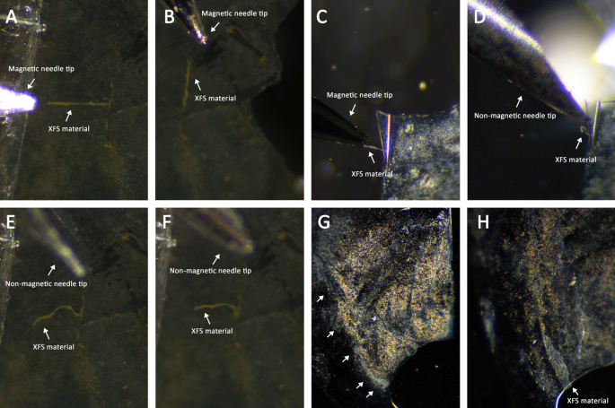
A magnetized pin was used to show that MP-bound XFS fibrils may very well be affected by way of a localized magnetic discipline. The magnetic drive generated on the tip of the pin was not sturdy sufficient to take away MP-XFS aggregates, it was capable of re-orient them within the route of the utilized discipline (Fig. 5A–C); opposite to a non-magnetic needle management (Fig. 5D–F). For some lens capsules these magnetic pins did begin to pull the perimeters of XFS supplies off the lens capsule, however was unable to take away these deposits completely (Fig. 5G, H). Nevertheless, any XFS supplies that had been already indifferent and in answer resulting from irrigation or surgical manipulation may very well be collected utilizing a magnetized pin.
Impact of magnetic pins on the magnetized XFS supplies. Human lens capsules having XFS supplies had been incubated with MP-peptide conjugates and subsequently, magnetic conduct of XFS aggregates was studied utilizing magnetic and non-magnetic instruments. (A, B) Giant aggregates of XFS supplies coated with MP-p-IPL had been pulled by magnetic pin in numerous instructions. (C, D) XFS supplies within the heart of lens capsule coated with MP-p-LPS had been drawn to the magnetic pin. Nevertheless, within the absence of a magnetic discipline (i.e. non-magnetic needle) no attraction was noticed. (E, F) Management research with non-magnetic needles confirmed that XFS supplies coated with MP-p-IPL didn’t react to the non-magnetic instrument. (G) XFS supplies within the heart of the human lens capsule coated with MP-peptide conjugates earlier than utility of magnetic pin. (H) Edges of the identical XFS deposits within the central zone of the lens capsule being pulled in direction of the utilized magnetic discipline by a magnetized pin
Rotating magnetic discipline research
Utilizing the identical experimental technique, a rotating magnetic discipline was employed to liberate XFS supplies from the lens capsule. It has been proven that the oscillation of disk-shaped MPs, by way of low-frequency rotating magnetic fields that produced a uniform 10,000 G magnetic discipline, might generate sufficient harmful drive to induce most cancers cell demise [21]. On this work, ex vivo research had been performed utilizing spherical MPs together with a 5000 G uniform magnetic discipline to generate mechanical drive for the removing of XFS deposits. Magnetic field-treated tissues had been then irrigated with BSS buffer to look at any attainable support in additional removing of XFS supplies from the tissues.
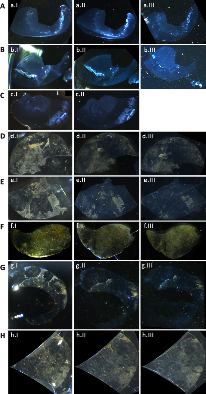
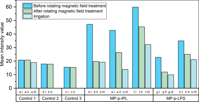
A rotating Halbach array was used to induce an exterior magnetic discipline on XFS laden lens capsules incubated with peptide-decorated MPs. It was noticed that the sector energy generated was adequate sufficient to result in the removing of a big quantity of XFS supplies from the floor of the lens capsule (Fig. 6). Moreover, the impact of irrigation after agitation by MPs below the magnetic discipline was used to judge the removing of those supplies. It was noticed that irrigation after magnetic discipline remedy result in an additional important removing of XFS supplies as in comparison with irrigation or MP remedy alone. For example, a lens capsule with central zone deposits that was certain to MP-p-IPL particles (Fig. 6D) confirmed a considerable amount of XFS removing upon making use of the rotating magnetic discipline (Fig. 6DII). One other tissue having XFS deposits on each central and peripheral zones (Fig. 6E), certain with MP-p-IPL particles, confirmed that the applying of the rotating magnetic discipline eliminated aggregates from each zones of the tissue (Fig. 6EII). MP-p-IPL certain supplies on a lens capsule with XFS deposits on the central zone had all massive XFS aggregates eliminated solely by the magnetic discipline (Fig. 6F). Nevertheless, this tissue had a dense quantity of cataractous supplies on the posterior facet of the lens capsule, which brought on the attachment of MPs to these supplies making the analysis tough. Due to this fact, though not as visually efficient because the earlier tissues, the rotating magnetic discipline was nonetheless truly efficient within the removing of the massive XFS aggregates from the floor of that lens capsule (Fig. 6FII). Lens capsules proven in (Fig. 6G, H)had been uncovered to MP-p-LPS conjugates and handled with the rotating magnetic discipline. The central zone of 1 pattern (Fig. 6G) was misplaced resulting from surgical procedure, nevertheless, XFS supplies on the lens floor had been principally eliminated after making use of the rotating magnetic discipline (Fig. 6GII). The rotating discipline was additionally efficient in eradicating a few of XFS aggregates from the opposite tissue (Fig. 6H) incubated with MP-p-LPS. On this case, the peripheral zone of the tissue was principally reduce out of the photographs as a result of coverslips used for holding the tissue in place throughout irrigation. Irrigation of BSS buffer over the tissue was proven to be efficient in eradicating some XFS aggregates from the floor of the lens capsule (Fig. 6D–H). The management tissues that had been handled with the rotating magnetic discipline confirmed nearly no removing of XFS supplies (Fig. 6A–C). As a result of tissue handing difficulties, we couldn’t do BSS irrigation on one of many management tissues (Fig. 6C). XFS supplies being partially misplaced within the management lens capsule proven in (Fig. 6BIII) was not as a result of impact of magnetic discipline, however relatively to pattern handing being answerable for the lack of the delicate supplies that had been already lifted from the lens capsule. The management lens capsule proven in (Fig. 6A) confirmed a little or no removing of XFS aggregates throughout BSS irrigation and no impact was noticed after treating with rotating magnetic discipline (Fig. 6AIII).
Impact of rotating magnetic discipline on XFS supplies. (A–C) Management XFS lens capsules handled with rotating magnetic discipline with out MPs. (D–F) XFS lens capsules interacted with MP-p-IPL. (G, H) XFS lens capsules interacted with MP-p-LPS. In every uncooked, pictures numbered as “I” symbolize tissues earlier than handled with rotating magnetic discipline, pictures numbered as “II” symbolize XFS lens capsule after 3 h remedy with rotating magnetic discipline, and pictures numbered as “III” symbolize lens capsules after buffer irrigation over the tissue
To higher consider the effectiveness of the noticed rotating magnetic discipline, pictures of tissues earlier than and after utilized magnetic discipline and irrigation (Fig. 6), had been analyzed primarily based on the depth of the lens capsule floor earlier than and after every take a look at (Fig. 7).
Imply depth measurements of XFS lens capsules incubated with MP-peptide conjugates. (Management 1-Management 3) Management tissues incubated with BSS and handled with rotating magnetic discipline. (MP-p-IPL) Consultant imply depth values measured utilizing pictures of XFS lens capsules interacted with MP-p-IPL. (MP-p-LPS) Imply depth values of various lens capsules interacted with MP-p-LPS. Each teams of graphs associated to check samples confirmed that intensities had been decreased as a result of removing of XFS supplies from the floor of lens capsules. In every graph, column “I” represents depth worth of the photographs of the floor of lens capsule earlier than treating with rotating magnetic discipline, column “II” represents values after 3 h remedy with the rotating magnet, and column “III” represents intensities after making use of BSS irrigation over the management XFS lens capsules. In all pictures, depth values have been reported after background subtraction
Cytotoxicity research of MP-peptide conjugates
Previous to toxicity research, the frequent phenotypic options of cultured hTM cells had been assessed utilizing immunohistochemical research (Further file 1: Fig. S1). The in vitro cytotoxicity of peptides in free and MP-conjugated kind was evaluated towards obtained hTM cells utilizing stay/useless cell viability assay, MTT assay, and DNA cleavage. All in vitro cell assays confirmed that each free peptide and MP-peptide particles weren’t poisonous within the concentrations studied, as detailed beneath.
Dwell/useless cell viability assay carried out on hTM monolayers
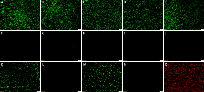
XFS-specific peptides in each answer free or MP certain kind had been incubated with hTM cells and their cytotoxic results assessed. It was discovered that each MP-p-IPL and MP-p-LPS constructs weren’t poisonous within the concentrations studied. Inexperienced represents stay cells and purple represents useless cells in (Fig. 8). Confluent cultured cells had been incubated with 50 µg and 100 µg of MP-p-IPL or MP-p-LPS (Fig. 8B–E). hTM monolayers had been additionally incubated with 1 mM of p-IPL or p-LPS peptide options, and their viability was analyzed in an analogous means because the MP-peptide constructs. As seen within the case of MP-peptide conjugates, free peptides weren’t poisonous in used concentrations (Fig. 8Okay, N) and Desk 1). Dwell/useless assay of hTM cells handled with rotating magnetic discipline additionally confirmed that cells incubated with MP-peptide conjugates didn’t present important loss in comparison with management cells incubated with water (Desk 2). Due to the sensitivity of hTM cells, it was not attainable to deal with cells with a rotating magnetic discipline exterior of a CO2 regulated ambiance for longer than 10 min. That stated, even management cells confirmed important loss upon removing from the CO2 incubator, thus, an optimum 10 min publicity to the magnetic discipline was chosen to judge the viability of cells within the presence of rotating magnetic discipline (Desk 2).
Cell viability (stay/useless) assay of hTM cells incubated with free peptide and MP-peptide constructs. Calcein AM (CAM)/ EthD-1 system: Calcein AM detects mobile esterase exercise of stay cells, EthD-1 stains nuclei of useless cells. The inexperienced cells are stay (CAM) and purple cells are useless cells (EthD-1). (A, F) Management cells handled with water (with out MPs). (B, G) hTM cells incubated with 50 µg MP-p-IPL for twenty-four h. (C, H) hTM cells incubated with 100 µg MP-p-IPL for twenty-four h. (D, I) hTM cells incubated with 50 µg MP-p-LPS for twenty-four h. (E, J) hTM cells incubated with 100 µg MP-p-IPL for twenty-four h. (Okay, L) hTM cells handled with 1 mM free p-IPL peptide answer (with out MPs). (M, N) hTM cells handled with 1 mM free p-LPS peptide answer (with out MPs). (O) All cells useless (methanol handled). Every remedy included three organic and three technical replicates. (Scales bars = 50 µm)
MTT cell proliferation assay
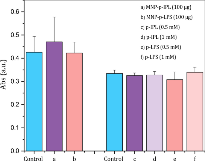
hTM cell proliferation within the presence of answer free and MP certain peptides was performed utilizing the MTT colorimetric assay. A one-way ANOVA take a look at confirmed that no important distinction (p < 0.05) was noticed between the absorbance of hTM cells handled with MP-peptide conjugates or answer free peptides in comparison with management cells handled with water (Fig. 9). The outcomes confirmed that each units of take a look at samples didn’t suppress the proliferation of hTM cells.
MTT colorimetric assay of viable hTM cells within the presence of XFS-specific peptides in free and conjugated kind. (Left) MTT assay performed utilizing peptide-conjugated MPs. (Proper) MTT assay outcomes after incubation of hTM cells for twenty-four h with 0.5 mM and 1 mM free peptide options. The optical density was measured at 570 nm after 24 h incubation of hTM cells with every take a look at pattern. (Knowledge symbolize imply ± 1 SD, n ≥ 3, no statistical distinction was noticed between the means)
DNA fragmentation
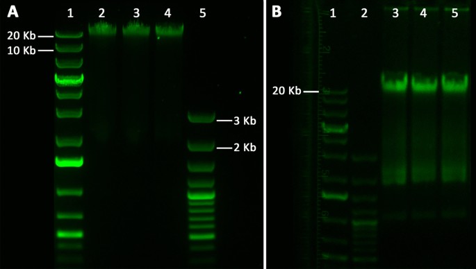
Impact of free and conjugated XFS-targeting peptides on the induction of apoptosis was investigated by way of chromosomal DNA fragmentation evaluation: DNA fragments point out apoptosis induction. MP-peptide constructs, in addition to answer free peptides, didn’t provoke apoptosis for hTM cells; chromosomal DNA remained intact after 24 h incubation with MP-p-IPL, MP-p-LPS, p-LPS, or p-IPL options (Fig. 10).
Electrophoretic analyses of the apoptotic chromosomal DNA fragmentation of hTM cells. (A) Remoted DNA from hTM cells after being incubated with MP-peptide conjugates for twenty-four h. lane 1: 1 Kb plus DNA ladder, lane 2: Management, hTM cells handled with water, lane 3: Extracted DNA of hTM cells incubated with 100 µg MP-p-IPL for twenty-four h, lane 4: Extracted DNA of hTM cells incubated with 100 µg MP-p-LPS for twenty-four h, lane 5: 100 bp plus DNA ladder. (B) Remoted DNA from hTM cells incubated with free XFS-targeting peptide options. Lane 1: 1 Kb plus DNA ladder, lane 2: 100 bp plus DNA ladder, lane 3: Management, hTM cells handled with water, lane 4: hTM cells incubated with 1 mM MP-p-IPL for twenty-four h, lane 4: hTM cells incubated with 1 mM MP-p-LPS for twenty-four h
Cell uptake research utilizing electron microscopy
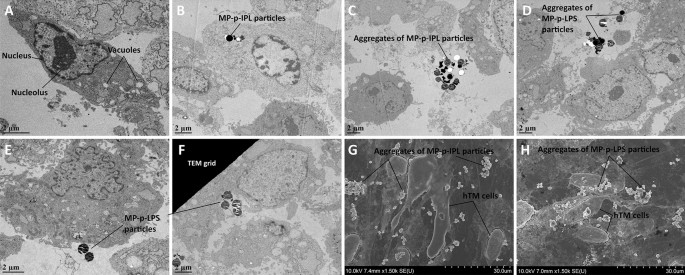
One of many key features of trabecular cells is the phagocytosis of international supplies and extracellular particles [33], a mobile perform additionally noticed in vitro [34]. The internalization of MP-peptide constructs into hTM cells might result in cell demise if uncovered to an oscillating magnetic discipline. Thus, hTM cells had been incubated for two h with 100 µg of every of MP-p-IPL or MP-p-LPS and TEM micrographs confirmed that particular person particles had been taken up by hTM cells (Fig. 11B), and a few particles had been engulfed by hTM cells (Fig. 11E, F). Aggregates of each MP-p-IPL and MP-p-LPS particles had been noticed exterior of cultured hTM cells as nicely (Fig. 11C, D). Particular person particles, in addition to aggregated MPs, had been additionally noticed in SEM micrographs of areas surrounding cultured hTM cells (Fig. 11G, H). Evidently MPs in massive aggregates have much less propensity to enter the classy cells. It has been beforehand proven that intravitreal or anterior chamber injection of magnetic nano- and microparticles had nearly no indicators of impact on retinal morphology, photoreceptor perform or IOP within the anterior chamber of animal fashions [35]. Regardless, based on completely different cell viability assays performed on this research, no statistical distinction was noticed for cell mortality upon incubation with MPs in cultured hTM cells.
Electron micrographs of hTM cells incubated with MP-peptide conjugates. (A–F) TEM micrographs. (G, H) SEM micrographs. hTM cells had been incubated with 100 µg of MP- peptide conjugates for two h. (A) Management hTM cells incubated with water. (B) MP-p-IPL particles positioned contained in the hTM cells. (C) Aggregates of MP-p-IPL particles positioned exterior of the cells. (D, E) Aggregates of MP-p-LPS particles positioned exterior of hTM cells. (F) MP-p-LPS particles being engulfed by the hTM cell. Black dust between particles may very well be trapped dye molecules. (G) SEM micrograph exhibiting aggregates of MP-p-IPL particles round hTM cells. (H) SEM micrograph of MP-p-LPS particles positioned across the hTM cells
[ad_2]


