
Metal Nanoparticles for Electrochemical Sensing: Progress and Challenges in the Clinical Transition of Point-of-Care Testing

 , ,
, ,
Abstract
:1. Introduction
2. Electrochemical Biosensors
2.1. Classification of ECBs Based on BRCs
- ECBs modified with biocatalysts
- ECBs operating through bioaffinity
2.1.1. ECBs Modified with Biocatalysts
2.1.2. ECBs Operating through Bioaffinity
- Immunosensors
- Aptasensors
- Affibody-based sensors
2.2. Classification of ECBs Based on EATs
- Amperometric technique
- Voltammetric technique
- Potentiometric technique
- Conductometric technique
- Impedimetric technique
2.2.1. Amperometric Technique
2.2.2. Voltammetric Technique
2.2.3. Potentiometric Technique
2.2.4. Conductometric Technique
2.2.5. Impedimetric Technique
3. MNPs and their Composites in ECBs
3.1. Influence of MNP Morphology in Biosensing
3.1.1. AuNPs: Effect of Size and Shape in Biosensing
3.1.2. AgNPs: Effect of Size and Shape in Biosensing
3.1.3. PtNPs: Effect of Size and Shape in Biosensing
3.2. Properties of MNPs Composites
3.2.1. Fullerene-Based MNPCs
3.2.2. CNT-Based MNPCs
3.2.3. GR-Based MNPCs
4. MNP-Based ECBs for Biomolecule Detection
4.1. MNPs in Small Biomolecule Sensing
4.2. MNPs in Cancer Biomarker Detection
4.2.1. MNPs in Carcinoma Embryonic Antigen Sensing
4.2.2. MNPs in Prostate-Specific Antigen Sensing
4.2.3. MNPs in Cancer Antigen 125 Sensing
4.2.4. MNPs in HER2 Sensing
4.2.5. MNPs in Alpha Fetoprotein Sensing
4.2.6. MNPs in Interleukin Sensing
4.3. MNPs in Novel Coronavirus Sensing
5. Advances in POCT Devices: Prospects and Challenges in the Clinical Transition of ECBs
- ECBs that utilize MNPs or MNPCs usually show higher sensitivity, stability, and wider linear range compared to those that do not utilize MNPs. However, it is important to choose which MNPs are most compatible with a specific BRC. Therefore, research into MNP and BRC compatibility might greatly enhance the stability of the fabricated ECBs.
- Using bi- or tri-metallic NPs significantly enhances the performance of ECBs. The possible reason for this is that the metal–metal interaction helps in lowering the HOMO-LUMO energy gap. This in turn allows for more active sites on the MNPs. Hence, using multimetallic NPs that interact synergistically with each other will allow for stronger interactions with the BRCs.
- MNPs with cubic, pyramidal, oval, and other unique shapes show higher catalytic activity and have an increased surface area compared to the commonly employed spherical MNPs. This would allow for immobilization of a greater amount of BRCs. At the same time, the edge sites of these MNPs show higher activity compared to the basal sites.
- MNPs that have QD or core shell-like structure show some unique physical, chemical, and electronic properties. These unique properties usually make them highly desirable for fabrication of ECBs.
- Green synthesis of MNPs is becoming ever popular. This not only allows for the preparation of MNPs in an environmentally friendly way, but also introduces various functional groups on the MNP surface. These functional groups, when properly utilized, might help in the robust anchoring of BRCs and enhance the stability and overall activity of the ECBs.
- MNPs interact differently with various CNMs. Hence, it is essential to properly choose the MNPs and CNMs before composites can be prepared for fabricating effective ECBs. Future research should focus on understanding the fundamental properties of various MNPCs. This would allow for the intelligent designing of MNPCs for fabricating ECBs.
- The screen-printing technique is most commonly used in the fabrication of ECB strips. However, methods such as inkjet printing, doctor blading, and aerosol-assisted chemical vapor deposition should be explored for determining the best approach for the fabrication of ECB strips.
- Aside from the above-mentioned topics, ECB researchers should work towards the commercialization of their laboratory models. This would then reveal the limitations of their proposed systems, and make way for future research that would help to overcome these shortcomings.
Funding
Acknowledgments
Conflicts of Interest
Abbreviations
| ADHD | Attention deficit hyperactivity disorder |
| AFP | Alpha-fetoprotein |
| Anb | Antibody |
| AgNPs | Silver NPs |
| Anb | Antibody |
| Ang | Antigen |
| AP | Auxiliary probe |
| APTES | 3-aminopropyltriethoxysilane |
| APTMS | 3-aminopropyltrimethoxysilane |
| ATPA | ATP aptamer |
| ASV | anodic stripping voltammetry |
| AuNPs | Gold NPs |
| BDD | Boron-doped diamond |
| BRC | biorecognition component |
| BSA | bovine serum albumin |
| CAS | Casein |
| CASPE-MFD | Commercially available screen-printed electrode-Based microfluidic devices |
| CDI | Carbonyldi-imidazole |
| cDNA | Complimentary DNA |
| CE | Counter electrode |
| CEA | Carcinoembryonic antigen |
| CNMs | Conducting nanomaterials |
| CNT | Carbon nanotube |
| conA | Concanavalin A |
| CP | Capture probe |
| CPE | Carbon paste electrode |
| CS | Chitosan |
| CSH | Copper silicate hollow spheres |
| CV | cyclic voltammetry |
| CysA | Cysteamine |
| DA | Dopamine |
| DMF | Dimethylformamide |
| DNR | Dendritic nanorods |
| DPV | differential pulse voltammetry |
| DPASV | differential pulse anodic stripping voltammetry |
| 3D-SP | 3D screen printed |
| EATs | electroanalytical techniques |
| ECBs | Electrochemical biosensors |
| ECD | Extracellular domain |
| EDC | N-(3,Dimethylaminopropyl)-N-ethyl-Carbodiimidehydrochloride |
| EIS | electrochemical impedance spectroscopy |
| ELISA | Enzyme-linked immunosorbent assay |
| ESR | Electron spin resonance |
| FA | Ferro-cenecarboxylic acid |
| FAD | Flavin adenosine dinucleotide |
| FC | Ferrocene |
| FLGR | few layer GR |
| fMWCNT | Functionalized MWCNTs |
| Glu | Glutaraldehyde |
| GNR | Gold nanorods |
| GNS | Gold Nanostructures |
| GO | GR oxide |
| GOx | glucose oxidase |
| GPTMS | Glycidyloxypropyl trimethoxysilane |
| GQD | Graphene quantum dot |
| GR | Graphene |
| GRNs | graphene nanoribbons |
| GRWs | GR nanowalls |
| HA | Histamine |
| HCC | Hepatocellular carcinoma |
| HepG2 | Hepatocellular carcinoma |
| HER2 | Human epidermal growth factor receptor 2 |
| HEV | Hepatitis E virus |
| HNF | Honey nanofibers |
| HNT | Halloysite nanotube |
| HP | Hairpin probe |
| HRP | horseradish peroxidase |
| HUA | Humic acid |
| IL | Interleukin |
| ISE | ion selective electrode |
| ITO | Indium tin oxide |
| LBL | Layer-by-layer |
| LIG | laser-induced graphene |
| LOD | limit of detection |
| LOx | Lactate oxidase |
| LP | Label probe |
| LPA-NHS | Lipoic acid N-hydroxysuccinimide ester |
| LSV | linear sweep voltammetry |
| 16-MUA | 16-Mercaptoundecanoic acid |
| MagB | Magnetic bead |
| MB | Methylene blue |
| MCH | 6-mercapto-1-hexanol |
| MLGR | multilayer GR |
| MNPs | Metal nanoparticles |
| MNPCs | MNP composites |
| MO | Methyl orange |
| MOF | Metal organic framework |
| MPA | 3-mercaptopropionic acid |
| MPTES | 3-mercaptopropyltriethoxysilane |
| MSA | Mercaptosuccinic acid |
| MWCNT | Multiwall CNT |
| 2019-nCoV | Novel coronavirus |
| NCMTs | Nitrogen-doped magnetic carbon microtubes |
| NF | Nanoflower |
| OC | Ovarian cancer |
| OHA | Ochratoxin A |
| pABA | p-aminobenzoic acid |
| PAMAM | Poly(amidoamine) |
| PAN-oxime | Amidoxime-modified polyacrylonitrile |
| PANI | Polyaniline |
| pATP | p-aminothiophenol |
| PB | Prussian blue |
| pCTAB | Poly cetyl trimethylammoniumbromide |
| PDA | Poly DA |
| PEDOT | Poly(3,4-ethylenedioxythiophene) |
| PEG | Polyethylene glycol |
| PGNR | porous graphene nanoribbon |
| PJI | Periprosthetic joint infection |
| PLA | Polylactic |
| POCT | Point-of-care testing |
| PGA | Poly(glutamic acid) |
| PPY | Polypyrrole |
| PS | Prostate cancer |
| PSA | Prostate specific antigen |
| PTK-7 | Protein tyrosine kinase-7 |
| PtNPs | Platinum NPs |
| PVA | Polyvinyl alcohol |
| RE | Reference electrode |
| rGO | Reduced GO |
| SBMs | Small biomolecules |
| SLGR | single layer GR |
| SPCE | Screen-printed carbon electrode |
| ssDNA | Single stranded DNA |
| ssRNA | Single stranded RNA |
| Strp | Streptavidin |
| SWCNT | Single wall CNT |
| SWV | square wave voltammetry |
| TB | Toluidine blue |
| TBA | Thrombin binding aptamer |
| TMV | tobacco mosaic virus |
| UA | Uric acid |
| UOx | Uricase |
| VBG | Vertical boron doped graphene |
| WE | Working electrode |
| XO | Xanthine oxidase |
References
- Ronkainen, N.J.; Halsall, H.B.; Heineman, W.R. Electrochemical biosensors. Chem. Soc. Rev. 2010, 39, 1747–1763. [Google Scholar] [CrossRef] [PubMed]
- Ouyang, M.; Tu, D.; Tong, L.; Sarwar, M.; Bhimaraj, A.; Li, C.; Coté, G.L.; Di Carlo, D. A review of biosensor technologies for blood biomarkers toward monitoring cardiovascular diseases at the point of care. Biosens. Bioelectron. 2020, 171, 112621. [Google Scholar] [CrossRef] [PubMed]
- Zhang, N.; Ruan, Y.F.; Zhang, L.B.; Zhao, W.W.; Xu, J.J.; Chen, H.Y. Nanochannels Photoelectrochemical Biosensor. Anal. Chem. 2018, 90, 2341–2347. [Google Scholar] [CrossRef] [PubMed]
- Chowdhury, A.D.; Takemura, K.; Li, T.C.; Suzuki, T.; Park, E.Y. Electrical pulse-induced electrochemical biosensor for hepatitis E virus detection. Nat. Commun. 2019, 10, 4–7. [Google Scholar] [CrossRef] [Green Version]
- Dai, Y.; Somoza, R.A.; Wang, L.; Welter, J.F.; Li, Y.; Caplan, A.I.; Liu, C.C. Exploring the Trans-Cleavage Activity of CRISPR-Cas12a (cpf1) for the Development of a Universal Electrochemical Biosensor. Angew. Chem. 2019, 131, 17560–17566. [Google Scholar] [CrossRef]
- Chen, L.C.; Wang, E.; Tai, C.S.; Chiu, Y.C.; Li, C.W.; Lin, Y.R.; Lee, T.H.; Huang, C.W.; Chen, J.C.; Chen, W.L. Improving the reproducibility, accuracy, and stability of an electrochemical biosensor platform for point-of-care use. Biosens. Bioelectron. 2020, 155, 112111. [Google Scholar] [CrossRef]
- Gill, A.A.S.; Singh, S.; Nate, Z.; Chauhan, R.; Thapliyal, N.B.; Karpoormath, R.; Maru, S.M.; Reddy, T.M. A novel copper-based 3D porous nanocomposite for electrochemical detection and inactivation of pathogenic bacteria. Sens. Actuators B Chem. 2020, 321, 128449. [Google Scholar] [CrossRef]
- Khan, M.Z.H.; Hasan, M.R.; Hossain, S.I.; Ahommed, M.S.; Daizy, M. Ultrasensitive detection of pathogenic viruses with electrochemical biosensor: State of the art. Biosens. Bioelectron. 2020, 166, 112431. [Google Scholar] [CrossRef]
- Ahammad, A.J.S.; Islam, T.; Hasan, M.M. Graphene-Based Electrochemical Sensors for Biomedical Applications. In Biomedical Applications of Graphene and 2D Nanomaterials; Elsevier Inc.: Amsterdam, The Netherlands, 2019; pp. 249–282. ISBN 9780128158890. [Google Scholar]
- Islam, T.; Hasan, M.M.; Akter, S.S.; Alharthi, N.H.; Karim, M.R.; Aziz, M.A.; Hossain, M.D.; Ahammad, A.J.S. Fabrication of Ni–Co-Based Heterometallo-Supramolecular Polymer Films and the Study of Electron Transfer Kinetics for the Nonenzymatic Electrochemical Detection of Nitrite. ACS Appl. Polym. Mater. 2020, 2, 273–284. [Google Scholar] [CrossRef]
- Chang, J.; Wang, X.; Wang, J.; Li, H.; Li, F. Nucleic Acid-Functionalized Metal-Organic Framework-Based Homogeneous Electrochemical Biosensor for Simultaneous Detection of Multiple Tumor Biomarkers. Anal. Chem. 2019, 91, 3604–3610. [Google Scholar] [CrossRef]
- Colozza, N.; Kehe, K.; Dionisi, G.; Popp, T.; Tsoutsoulopoulos, A.; Steinritz, D.; Moscone, D.; Arduini, F. A wearable origami-like paper-based electrochemical biosensor for sulfur mustard detection. Biosens. Bioelectron. 2019, 129, 15–23. [Google Scholar] [CrossRef] [PubMed]
- Ahammad, A.J.S.; Alam, M.K.; Islam, T.; Hasan, M.M.; Karim, R.; Anju, A.N.; Mozumder, M.N.I. Poly (brilliant cresyl blue)-reduced graphene oxide modified activated GCE for nitrite detection: Analyzing the synergistic interactions through experimental and computational study. Electrochim. Acta 2020, 349, 136375. [Google Scholar] [CrossRef]
- Zhou, L.; Wang, Y.; Yang, C.; Xu, H.; Luo, J.; Zhang, W.; Tang, X.; Yang, S.; Fu, W.; Chang, K.; et al. A label-free electrochemical biosensor for microRNAs detection based on DNA nanomaterial by coupling with Y-shaped DNA structure and non-linear hybridization chain reaction. Biosens. Bioelectron. 2019, 126, 657–663. [Google Scholar] [CrossRef] [PubMed]
- Sun, A.C.; Hall, D.A. Point-of-Care Smartphone-based Electrochemical Biosensing. Electroanalysis 2019, 31, 2–16. [Google Scholar] [CrossRef] [Green Version]
- Haque, M.A.; Hasan, M.M.; Islam, T.; Razzak, M.A.; Alharthi, N.H.; Sindan, A.; Karim, M.R.; Basha, S.I.; Aziz, M.A.; Ahammad, A.J.S. Hollow Reticular Shaped Highly Ordered Rice Husk Carbon for the Simultaneous Determination of Dopamine and Uric Acid. Electroanalysis 2020, 1–15. [Google Scholar] [CrossRef]
- Chellapandian, C.; Ramkumar, B.; Puja, P.; Shanmuganathan, R.; Pugazhendhi, A.; Kumar, P. Gold nanoparticles using red seaweed Gracilaria verrucosa: Green synthesis, characterization and biocompatibility studies. Process Biochem. 2019, 80, 58–63. [Google Scholar] [CrossRef]
- Kang, M.S.; Lee, S.Y.; Kim, K.S.; Han, D.W. State of the art biocompatible gold nanoparticles for cancer theragnosis. Pharmaceutics 2020, 12, 701. [Google Scholar] [CrossRef] [PubMed]
- Baygar, T.; Sarac, N.; Ugur, A.; Karaca, I.R. Antimicrobial characteristics and biocompatibility of the surgical sutures coated with biosynthesized silver nanoparticles. Bioorg. Chem. 2019, 86, 254–258. [Google Scholar] [CrossRef]
- Eanest Jebasingh, B.; Valan Arasu, A. A comprehensive review on latent heat and thermal conductivity of nanoparticle dispersed phase change material for low-temperature applications. Energy Storage Mater. 2020, 24, 52–74. [Google Scholar] [CrossRef]
- Agarwal, H.; Nakara, A.; Shanmugam, V.K. Anti-inflammatory mechanism of various metal and metal oxide nanoparticles synthesized using plant extracts: A review. Biomed. Pharmacother. 2019, 109, 2561–2572. [Google Scholar] [CrossRef]
- Nurunnabi, M.; Nafiujjaman, M.; Lee, S.J.; Park, I.K.; Huh, K.M.; Lee, Y.K. Preparation of ultra-thin hexagonal boron nitride nanoplates for cancer cell imaging and neurotransmitter sensing. Chem. Commun. 2016, 52, 6146–6149. [Google Scholar] [CrossRef] [PubMed]
- Han, S.; Liu, W.; Zheng, M.; Wang, R. Label-Free and Ultrasensitive Electrochemical DNA Biosensor Based on Urchinlike Carbon Nanotube-Gold Nanoparticle Nanoclusters. Anal. Chem. 2020, 92, 4780–4787. [Google Scholar] [CrossRef] [PubMed]
- Lv, Q.; Wang, Y.; Su, C.; Lakshmipriya, T.; Gopinath, S.C.B.; Pandian, K.; Perumal, V.; Liu, Y. Human papilloma virus DNA-biomarker analysis for cervical cancer: Signal enhancement by gold nanoparticle-coupled tetravalent streptavidin-biotin strategy. Int. J. Biol. Macromol. 2019, 134, 354–360. [Google Scholar] [CrossRef] [PubMed]
- Yuan, Q.; Liu, Y.; Ye, C.; Sun, H.; Dai, D.; Wei, Q.; Lai, G.; Wu, T.; Yu, A.; Fu, L.; et al. Highly stable and regenerative graphene–diamond hybrid electrochemical biosensor for fouling target dopamine detection. Biosens. Bioelectron. 2018, 111, 117–123. [Google Scholar] [CrossRef] [PubMed]
- Xu, S.; Huang, X.; Chen, Y.; Liu, Y.; Zhao, W.; Sun, Z.; Zhu, Y.; Liu, X.; Wong, C.P. Silver Nanoparticle-Enzyme Composite Films for Hydrogen Peroxide Detection. ACS Appl. Nano Mater. 2019, 2, 5910–5921. [Google Scholar] [CrossRef]
- Nguyen, T.N.H.; Nolan, J.K.; Park, H.; Lam, S.; Fattah, M.; Page, J.C.; Joe, H.E.; Jun, M.B.G.; Lee, H.; Kim, S.J.; et al. Facile fabrication of flexible glutamate biosensor using direct writing of platinum nanoparticle-based nanocomposite ink. Biosens. Bioelectron. 2019, 131, 257–266. [Google Scholar] [CrossRef]
- Parate, K.; Karunakaran, C.; Claussen, J.C. Electrochemical cotinine sensing with a molecularly imprinted polymer on a graphene-platinum nanoparticle modified carbon electrode towards cigarette smoke exposure monitoring. Sens. Actuators B Chem. 2019, 287, 165–172. [Google Scholar] [CrossRef]
- Hosseini, G.M.; Mirzaei, M.; Torkzadeh-Mahani, M. Electrochemical aptasensor for activated protein C using a gold nanoparticle—Chitosan/graphene paste modified carbon paste electrode. Bioelectrochemistry 2019, 130, 107322. [Google Scholar] [CrossRef]
- Hou, L.; Huang, Y.; Hou, W.; Yan, Y.; Liu, J.; Xia, N. Modification-free amperometric biosensor for the detection of wild-type p53 protein based on the in situ formation of silver nanoparticle networks for signal amplification. Int. J. Biol. Macromol. 2020, 158, 580–586. [Google Scholar] [CrossRef]
- Wang, L.; Hao, L.; Qi, W.; Huo, X.; Xue, L.; Liu, Y.; Zhang, Q.; Lin, J. A sensitive Salmonella biosensor using platinum nanoparticle loaded manganese dioxide nanoflowers and thin-film pressure detector. Sens. Actuators B Chem. 2020, 321, 128616. [Google Scholar] [CrossRef]
- Roach, K.A.; Stefaniak, A.B.; Roberts, J.R. Metal nanomaterials: Immune effects and implications of physicochemical properties on sensitization, elicitation, and exacerbation of allergic disease. J. Immunotoxicol. 2019, 16, 87–124. [Google Scholar] [CrossRef]
- Jamkhande, P.G.; Ghule, N.W.; Bamer, A.H.; Kalaskar, M.G. Metal nanoparticles synthesis: An overview on methods of preparation, advantages and disadvantages, and applications. J. Drug Deliv. Sci. Technol. 2019, 53, 101174. [Google Scholar] [CrossRef]
- Gherab, K.; AI-Douri, Y.; Hashim, U.; Ameri, M.; Bouhemadou, A.; Batoo, K.M.; Adil, S.F.; Khan, M.; Raslan, E.H. Fabrication and characterizations of Al nanoparticles doped ZnO nanostructures-based integrated electrochemical biosensor. J. Mater. Res. Technol. 2020, 9, 857–867. [Google Scholar] [CrossRef]
- Asadian, E.; Ghalkhani, M.; Shahrokhian, S. Sensors and Actuators B: Chemical Electrochemical sensing based on carbon nanoparticles: A review. Sens. Actuators B. Chem. 2019, 293, 183–209. [Google Scholar] [CrossRef]
- Shetti, N.P.; Bukkitgar, S.D.; Reddy, K.R.; Reddy, C.V.; Aminabhavi, T.M. Nanostructured titanium oxide hybrids-based electrochemical biosensors for healthcare applications. Colloids Surf. B Biointerfaces 2019, 178, 385–394. [Google Scholar] [CrossRef] [PubMed]
- Zhu, L.; Miao, M.; Shao, X.; Du, Z.; Huang, K.; Luo, Y.; Xu, W. A Universal Electrochemical Biosensor Using Nick-HCR Nanostructure as Molecular Gate of Nanochannel for Detecting Chromium(III) Ions and MicroRNA. Anal. Chem. 2019, 91, 14992–14999. [Google Scholar] [CrossRef]
- Dimcheva, N. Nanostructures of noble metals as functional materials in biosensors. Curr. Opin. Electrochem. 2020, 19, 35–41. [Google Scholar] [CrossRef]
- Chen, X.; Huang, J.; Zhang, S.; Mo, F.; Su, S.; Li, Y.; Fang, L.; Deng, J.; Huang, H.; Luo, Z.; et al. Electrochemical Biosensor for DNA Methylation Detection through Hybridization Chain-Amplified Reaction Coupled with a Tetrahedral DNA Nanostructure. ACS Appl. Mater. Interfaces 2019, 11, 3745–3752. [Google Scholar] [CrossRef] [PubMed]
- Li, Z.; Wang, L.; Li, Y.; Feng, Y.; Feng, W. Carbon-based functional nanomaterials: Preparation, properties and applications. Compos. Sci. Technol. 2019, 179, 10–40. [Google Scholar] [CrossRef]
- Du, X.; Zhang, Z.; Zheng, X.; Zhang, H.; Dong, D.; Zhang, Z.; Liu, M.; Zhou, J. An electrochemical biosensor for the detection of epithelial-mesenchymal transition. Nat. Commun. 2020, 11, 192. [Google Scholar] [CrossRef] [Green Version]
- Ayaz, S.; Karakaya, S.; Emir, G.; Dilgin, D.G.; Dilgin, Y. A novel enzyme-free FI-amperometric glucose biosensor at Cu nanoparticles modified graphite pencil electrode. Microchem. J. 2020, 154, 104586. [Google Scholar] [CrossRef]
- Hondred, J.A.; Medintz, I.L.; Claussen, J.C. Enhanced electrochemical biosensor and supercapacitor with 3D porous architectured graphene via salt impregnated inkjet maskless lithography. Nanoscale Horiz. 2019, 4, 735–746. [Google Scholar] [CrossRef] [Green Version]
- Baek, S.H.; Roh, J.; Park, C.Y.; Kim, M.W.; Shi, R.; Kailasa, S.K.; Park, T.J. Cu-nanoflower decorated gold nanoparticles-graphene oxide nanofiber as electrochemical biosensor for glucose detection. Mater. Sci. Eng. C 2020, 107, 110273. [Google Scholar] [CrossRef] [PubMed]
- Lu, J.; Hu, Y.; Wang, P.; Liu, P.; Chen, Z.; Sun, D. Electrochemical biosensor based on gold nanoflowers-encapsulated magnetic metal-organic framework nanozymes for drug evaluation with in-situ monitoring of H2O2 released from H9C2 cardiac cells. Sens. Actuators B Chem. 2020, 311, 127909. [Google Scholar] [CrossRef]
- Gattani, A.; Singh, S.V.; Agrawal, A.; Khan, M.H.; Singh, P. Recent progress in electrochemical biosensors as point of care diagnostics in livestock health. Anal. Biochem. 2019, 579, 25–34. [Google Scholar] [CrossRef]
- Mohanraj, J.; Durgalakshmi, D.; Rakkesh, R.A.; Balakumar, S.; Rajendran, S.; Karimi-Maleh, H. Facile synthesis of paper based graphene electrodes for point of care devices: A double stranded DNA (dsDNA) biosensor. J. Colloid Interface Sci. 2020, 566, 463–472. [Google Scholar] [CrossRef] [PubMed]
- Newman, J.D.; Turner, A.P.F. Home blood glucose biosensors: A commercial perspective. Biosens. Bioelectron. 2005, 20, 2435–2453. [Google Scholar] [CrossRef] [Green Version]
- Zhao, C.; Thuo, M.M.; Liu, X. Erratum: A microfluidic paper-based electrochemical biosensor array for multiplexed detection of metabolic biomarkers (Science and Technology of Advanced Materials (2013) 14 (054402)). Sci. Technol. Adv. Mater. 2015, 16, 054402. [Google Scholar] [CrossRef]
- Liu, J.; Siavash Moakhar, R.; Sudalaiyadum Perumal, A.; Roman, H.N.; Mahshid, S.; Wachsmann-Hogiu, S. An AgNP-deposited commercial electrochemistry test strip as a platform for urea detection. Sci. Rep. 2020, 10, 9527. [Google Scholar] [CrossRef]
- Loncaric, C.; Tang, Y.; Ho, C.; Parameswaran, M.A.; Yu, H.Z. A USB-based electrochemical biosensor prototype for point-of-care diagnosis. Sens. Actuators B Chem. 2012, 161, 908–913. [Google Scholar] [CrossRef]
- Uniyal, S.; Sharma, R.K. Technological advancement in electrochemical biosensor based detection of Organophosphate pesticide chlorpyrifos in the environment: A review of status and prospects. Biosens. Bioelectron. 2018, 116, 37–50. [Google Scholar] [CrossRef] [PubMed]
- Shetti, N.P.; Malode, S.J.; Roy, S.; Chandra, P.; Reddy, K.R.; Chatterjee, S. Electroanalytical techniques for investigating biofilms: Applications in biosensing and biomolecular interfacing. In Nanomaterials in Diagnostic Tools and Devices; Elsevier: Amsterdam, The Netherlands, 2020; ISBN 9780128179239. [Google Scholar]
- Farka, Z.; Juřík, T.; Kovář, D.; Trnková, L.; Skládal, P. Nanoparticle-Based Immunochemical Biosensors and Assays: Recent Advances and Challenges. Chem. Rev. 2017, 117, 9973–10042. [Google Scholar] [CrossRef] [PubMed]
- Hasan, A.; Nurunnabi, M.; Morshed, M.; Paul, A.; Polini, A.; Kuila, T.; Al Hariri, M.; Lee, Y.K.; Jaffa, A.A. Recent advances in application of biosensors in tissue engineering. Biomed Res. Int. 2014, 2014. [Google Scholar] [CrossRef] [PubMed] [Green Version]
- Cipolatti, E.P.; Valério, A.; Henriques, R.O.; Moritz, D.E.; Ninow, J.L.; Freire, D.M.G.; Manoel, E.A.; Fernandez-Lafuente, R.; De Oliveira, D. Nanomaterials for biocatalyst immobilization-state of the art and future trends. RSC Adv. 2016, 6, 104675–104692. [Google Scholar] [CrossRef]
- Bilal, M.; Adeel, M.; Rasheed, T.; Iqbal, H.M.N. Multifunctional metal-organic frameworks-based biocatalytic platforms: Recent developments and future prospects. J. Mater. Res. Technol. 2019, 8, 2359–2371. [Google Scholar] [CrossRef]
- Hollmann, F.; Schmid, A. Electrochemical regeneration of oxidoreductases for cell-free biocatalytic redox reactions. Biocatal. Biotransform. 2004, 22, 63–88. [Google Scholar] [CrossRef]
- Bandodkar, A.J.; Jia, W.; Ramírez, J.; Wang, J. Biocompatible Enzymatic Roller Pens for Direct Writing of Biocatalytic Materials: “Do-it-Yourself” Electrochemical Biosensors. Adv. Healthc. Mater. 2015, 4, 1215–1224. [Google Scholar] [CrossRef]
- Campaña, A.L.; Florez, S.L.; Noguera, M.J.; Fuentes, O.P.; Puentes, P.R.; Cruz, J.C.; Osma, J.F. Enzyme-based electrochemical biosensors for microfluidic platforms to detect pharmaceutical residues in wastewater. Biosensors 2019, 9, 41. [Google Scholar] [CrossRef] [Green Version]
- Kucherenko, I.S.; Soldatkin, O.O.; Dzyadevych, S.V.; Soldatkin, A.P. Electrochemical biosensors based on multienzyme systems: Main groups, advantages and limitations—A review. Anal. Chim. Acta 2020, 1111, 114–131. [Google Scholar] [CrossRef]
- López Marzo, A.M.; Mayorga-Martinez, C.C.; Pumera, M. 3D-printed graphene direct electron transfer enzyme biosensors. Biosens. Bioelectron. 2020, 151, 111980. [Google Scholar] [CrossRef]
- Bäcker, M.; Koch, C.; Eiben, S.; Geiger, F.; Eber, F.; Gliemann, H.; Poghossian, A.; Wege, C.; Schöning, M.J. Tobacco mosaic virus as enzyme nanocarrier for electrochemical biosensors. Sens. Actuators B Chem. 2017, 238, 716–722. [Google Scholar] [CrossRef]
- Alizadeh, N.; Salimi, A. Ultrasensitive Bioaffinity Electrochemical Sensors: Advances and New Perspectives. Electroanalysis 2018, 30, 2803–2840. [Google Scholar] [CrossRef]
- Tu, J.; Torrente-Rodríguez, R.M.; Wang, M.; Gao, W. The Era of Digital Health: A Review of Portable and Wearable Affinity Biosensors. Adv. Funct. Mater. 2020, 30, 1906713. [Google Scholar] [CrossRef]
- Huang, Y.; Xu, J.; Liu, J.; Wang, X.; Chen, B. Disease-related detection with electrochemical biosensors: A review. Sensors 2017, 17, 2375. [Google Scholar] [CrossRef]
- Abdorahim, M.; Rabiee, M.; Alhosseini, S.N.; Tahriri, M.; Yazdanpanah, S.; Alavi, S.H.; Tayebi, L. Nanomaterials-based electrochemical immunosensors for cardiac troponin recognition: An illustrated review. TrAC Trends Anal. Chem. 2016, 82, 337–347. [Google Scholar] [CrossRef]
- Cho, I.H.; Lee, J.; Kim, J.; Kang, M.S.; Paik, J.K.; Ku, S.; Cho, H.M.; Irudayaraj, J.; Kim, D.H. Current technologies of electrochemical immunosensors: Perspective on signal amplification. Sensors 2018, 18, 207. [Google Scholar] [CrossRef] [Green Version]
- Khanmohammadi, A.; Aghaie, A.; Vahedi, E.; Qazvini, A.; Ghanei, M.; Afkhami, A.; Hajian, A.; Bagheri, H. Electrochemical biosensors for the detection of lung cancer biomarkers: A review. Talanta 2020, 206, 120251. [Google Scholar] [CrossRef]
- Carneiro, P.; Loureiro, J.; Delerue-Matos, C.; Morais, S.; do Carmo Pereira, M. Alzheimer’s disease: Development of a sensitive label-free electrochemical immunosensor for detection of amyloid beta peptide. Sens. Actuators B Chem. 2017, 239, 157–165. [Google Scholar] [CrossRef] [Green Version]
- de Castro, A.C.H.; Alves, L.M.; Siquieroli, A.C.S.; Madurro, J.M.; Brito-Madurro, A.G. Label-free electrochemical immunosensor for detection of oncomarker CA125 in serum. Microchem. J. 2020, 155, 104746. [Google Scholar] [CrossRef]
- Camilo, D.E.; Miyazaki, C.M.; Shimizu, F.M.; Ferreira, M. Improving direct immunoassay response by layer-by-layer films of gold nanoparticles—Antibody conjugate towards label-free detection. Mater. Sci. Eng. C 2019, 102, 315–323. [Google Scholar] [CrossRef]
- Gogola, J.L.; Martins, G.; Caetano, F.R.; Ricciardi-Jorge, T.; Duarte dos Santos, C.N.; Marcolino-Junior, L.H.; Bergamini, M.F. Label-free electrochemical immunosensor for quick detection of anti-hantavirus antibody. J. Electroanal. Chem. 2019, 842, 140–145. [Google Scholar] [CrossRef]
- Filik, H.; Avan, A.A. Nanostructures for nonlabeled and labeled electrochemical immunosensors: Simultaneous electrochemical detection of cancer markers: A review. Talanta 2019, 205, 120153. [Google Scholar] [CrossRef] [PubMed]
- Pan, G.; Zhao, G.; Wei, M.; Wang, Y.; Zhao, B. Design of nanogold electrochemical immunosensor for detection of four phenolic estrogens. Chem. Phys. Lett. 2019, 732, 136657. [Google Scholar] [CrossRef]
- Jampasa, S.; Pummoree, J.; Siangproh, W.; Khongchareonporn, N. Sensors and Actuators B: Chemical “ Signal-On ” electrochemical biosensor based on a competitive immunoassay format for the sensitive determination of oxytetracycline. Sens. Actuators B. Chem. 2020, 320, 128389. [Google Scholar] [CrossRef]
- Hou, L.; Zhang, X.; Kong, M.; Jiang, G.; Sun, Y.; Mo, W.; Lin, T.; Ye, F.; Zhao, S. A competitive immunoassay for electrochemical impedimetric determination of chlorpyrifos using a nanogold-modified glassy carbon electrode based on enzymatic biocatalytic precipitation. Microchim. Acta 2020, 187, 204. [Google Scholar] [CrossRef]
- Pei, X.; Zhang, B.; Tang, J.; Liu, B.; Lai, W.; Tang, D. Sandwich-type immunosensors and immunoassays exploiting nanostructure labels: A review. Anal. Chim. Acta 2013, 758, 1–18. [Google Scholar] [CrossRef]
- Felix, F.S.; Angnes, L. Electrochemical immunosensors—A powerful tool for analytical applications. Biosens. Bioelectron. 2018, 102, 470–478. [Google Scholar] [CrossRef]
- Jampasa, S.; Lae-ngee, P.; Patarakul, K.; Ngamrojanavanich, N.; Chailapakul, O.; Rodthongkum, N. Electrochemical immunosensor based on gold-labeled monoclonal anti-LipL32 for leptospirosis diagnosis. Biosens. Bioelectron. 2019, 142, 111539. [Google Scholar] [CrossRef]
- Mishra, G.K.; Sharma, V.; Mishra, R.K. Electrochemical aptasensors for food and environmental safeguarding: A review. Biosensors 2018, 8, 28. [Google Scholar] [CrossRef] [Green Version]
- Yoon, J.-Y. Basic Principles of Electrochemical Biosensing Using a Smartphone; Elsevier Inc.: Amsterdam, The Netherlands, 2020; ISBN 9780128170441. [Google Scholar]
- Shrivastava, S.; Trung, T.Q.; Lee, N.E. Recent progress, challenges, and prospects of fully integrated mobile and wearable point-of-care testing systems for self-testing. Chem. Soc. Rev. 2020, 49, 1812–1866. [Google Scholar] [CrossRef]
- Yao, Y.; Jiang, C.; Ping, J. Flexible freestanding graphene paper-based potentiometric enzymatic aptasensor for ultrasensitive wireless detection of kanamycin. Biosens. Bioelectron. 2019, 123, 178–184. [Google Scholar] [CrossRef] [PubMed]
- Dunn, M.R.; Mccloskey, C.M.; Buckley, P.; Rhea, K.; Chaput, J.C. Generating Biologically Stable TNA Aptamers that Function with High Affinity and Thermal Stability. J. Am. Chem. Soc. 2020, 142, 7721–7724. [Google Scholar] [CrossRef] [PubMed]
- Su, S.; Sun, H.; Cao, W.; Chao, J.; Peng, H.; Zuo, X.; Yuwen, L.; Fan, C.; Wang, L. Dual-Target Electrochemical Biosensing Based on DNA Structural Switching on Gold Nanoparticle-Decorated MoS2 Nanosheets. ACS Appl. Mater. Interfaces 2016, 8, 6826–6833. [Google Scholar] [CrossRef] [PubMed]
- Wang, L.; Wen, Y.; Yang, X.; Xu, L.; Liang, W.; Zhu, Y.; Wang, L.; Li, Y.; Li, Y.; Ding, M.; et al. Ultrasensitive Electrochemical DNA Biosensor Based on a Label-Free Assembling Strategy Using a Triblock polyA DNA Probe. Anal. Chem. 2019, 91, 16002–16009. [Google Scholar] [CrossRef] [PubMed]
- Mazaafrianto, D.N.; Ishida, A.; Maeki, M.; Tani, H.; Tokeshi, M. An electrochemical sensor based on structure switching of dithiol-modified aptamer for simple detection of Ochratoxin A. Anal. Sci. 2019, 35, 1221–1226. [Google Scholar] [CrossRef] [PubMed] [Green Version]
- Wang, Q.L.; Cui, H.F.; Song, X.; Fan, S.F.; Chen, L.L.; Li, M.M.; Li, Z.Y. A label-free and lectin-based sandwich aptasensor for detection of carcinoembryonic antigen. Sens. Actuators B Chem. 2018, 260, 48–54. [Google Scholar] [CrossRef]
- Abnous, K.; Danesh, N.M.; Ramezani, M.; Emrani, A.S.; Taghdisi, S.M. A novel colorimetric sandwich aptasensor based on an indirect competitive enzyme-free method for ultrasensitive detection of chloramphenicol. Biosens. Bioelectron. 2016, 78, 80–86. [Google Scholar] [CrossRef]
- Zhong, H.; Yu, C.; Gao, R.; Chen, J.; Yu, Y.; Geng, Y.; Wen, Y.; He, J. A novel sandwich aptasensor for detecting T-2 toxin based on rGO-TEPA-Au@Pt nanorods with a dual signal amplification strategy. Biosens. Bioelectron. 2019, 144, 111635. [Google Scholar] [CrossRef]
- Farzin, L.; Sadjadi, S.; Shamsipur, M.; Sheibani, S.; Mousazadeh, M. hasan Employing AgNPs doped amidoxime-modified polyacrylonitrile (PAN-oxime) nanofibers for target induced strand displacement-based electrochemical aptasensing of CA125 in ovarian cancer patients. Mater. Sci. Eng. C 2019, 97, 679–687. [Google Scholar] [CrossRef]
- Li, Z.; Zhou, Z.; Xue, N.; Wu, S.; Miao, X. Electrochemical aptamer-based determination of protein tyrosine kinase-7 using toehold-mediated strand displacement amplification on gold nanoparticles and graphene oxide. Microchim. Acta 2019, 186, 720. [Google Scholar] [CrossRef]
- Wang, Y.; Yao, L.; Ning, G.; Wu, Y.; Wu, S.; Mao, S.; Liu, G.Q. An electrochemical strategy for tetracycline detection coupled triple helix aptamer probe with catalyzed hairpin assembly signal amplification. Biosens. Bioelectron. 2019, 143, 111613. [Google Scholar] [CrossRef] [PubMed]
- Nameghi, M.A.; Danesh, N.M.; Ramezani, M.; Alibolandi, M.; Abnous, K.; Taghdisi, S.M. An ultrasensitive electrochemical sensor for 17β-estradiol using split aptamers. Anal. Chim. Acta 2019, 1065, 107–112. [Google Scholar] [CrossRef] [PubMed]
- Jiang, Y.; Sun, D.; Liang, Z.; Chen, L.; Zhang, Y.; Chen, Z. Label-free and competitive aptamer cytosensor based on layer-by-layer assembly of DNA-platinum nanoparticles for ultrasensitive determination of tumor cells. Sens. Actuators B Chem. 2018, 262, 35–43. [Google Scholar] [CrossRef]
- Cui, F.; Zhou, Z.; Zhou, H.S. Review—Measurement and Analysis of Cancer Biomarkers Based on Electrochemical Biosensors. J. Electrochem. Soc. 2020, 167, 037525. [Google Scholar] [CrossRef]
- Löfblom, J.; Feldwisch, J.; Tolmachev, V.; Carlsson, J.; Ståhl, S.; Frejd, F.Y. Affibody molecules: Engineered proteins for therapeutic, diagnostic and biotechnological applications. FEBS Lett. 2010, 584, 2670–2680. [Google Scholar] [CrossRef] [PubMed] [Green Version]
- Frejd, F.Y.; Kim, K.T. Affibody molecules as engineered protein drugs. Exp. Mol. Med. 2017, 49, e306. [Google Scholar] [CrossRef] [PubMed] [Green Version]
- Ravalli, A.; da Rocha, C.G.; Yamanaka, H.; Marrazza, G. A label-free electrochemical affisensor for cancer marker detection: The case of HER2. Bioelectrochemistry 2015, 106, 268–275. [Google Scholar] [CrossRef] [PubMed]
- Tang, Z.; Ma, Z. Multiple functional strategies for amplifying sensitivity of amperometric immunoassay for tumor markers: A review. Biosens. Bioelectron. 2017, 98, 100–112. [Google Scholar] [CrossRef]
- Ahammad, A.J.S.; Pal, P.R.; Shah, S.S.; Islam, T.; Hasan, M.M.; Qasem, M.A.A.; Odhikari, N.; Sarker, S.; Kim, D.M.; Aziz, M.A. Activated jute carbon paste screen-printed FTO electrodes for nonenzymatic amperometric determination of nitrite. J. Electroanal. Chem. 2019, 832, 368–379. [Google Scholar] [CrossRef]
- Joshi, A.; Kim, K. Biosensors and Bioelectronics Recent advances in nanomaterial-based electrochemical detection of antibiotics: Challenges and future perspectives. Biosens. Bioelectron. 2020, 153, 112046. [Google Scholar] [CrossRef]
- Sahin, B.; Kaya, T. Electrochemical amperometric biosensor applications of nanostructured metal oxides: A review. Mater. Res. Express 2019, 6, 042003. [Google Scholar] [CrossRef]
- Dong, X.X.; Yang, J.Y.; Luo, L.; Zhang, Y.F.; Mao, C.; Sun, Y.M.; Lei, H.T.; Shen, Y.D.; Beier, R.C.; Xu, Z.L. Portable amperometric immunosensor for histamine detection using Prussian blue-chitosan-gold nanoparticle nanocomposite films. Biosens. Bioelectron. 2017, 98, 305–309. [Google Scholar] [CrossRef] [PubMed] [Green Version]
- Muñoz, J.; Montes, R.; Bastos-Arrieta, J.; Guardingo, M.; Busqué, F.; Ruíz-Molina, D.; Palet, C.; García-Orellana, J.; Baeza, M. Carbon nanotube-based nanocomposite sensor tuned with a catechol as novel electrochemical recognition platform of uranyl ion in aqueous samples. Sens. Actuators B Chem. 2018, 273, 1807–1815. [Google Scholar] [CrossRef]
- Meirinho, S.G.; Dias, L.G.; Peres, A.M.; Rodrigues, L.R. Voltammetric aptasensors for protein disease biomarkers detection: A review. Biotechnol. Adv. 2016, 34, 941–953. [Google Scholar] [CrossRef] [PubMed] [Green Version]
- Shrivastava, S.; Jadon, N.; Jain, R. Next-generation polymer nanocomposite-based electrochemical sensors and biosensors: A review. TrAC Trends Anal. Chem. 2016, 82, 55–67. [Google Scholar] [CrossRef]
- Kumar, T.H.V.; Sundramoorthy, A.K. Electrochemical biosensor for methyl parathion based on single-walled carbon nanotube/glutaraldehyde crosslinked acetylcholinesterase-wrapped bovine serum albumin nanocomposites. Anal. Chim. Acta 2019, 1074, 131–141. [Google Scholar] [CrossRef]
- Diouf, A.; Motia, S.; El Alami El Hassani, N.; El Bari, N.; Bouchikhi, B. Development and characterization of an electrochemical biosensor for creatinine detection in human urine based on functional molecularly imprinted polymer. J. Electroanal. Chem. 2017, 788, 44–53. [Google Scholar] [CrossRef]
- Scholz, F. Voltammetric techniques of analysis: The essentials. ChemTexts 2015, 1, 17. [Google Scholar] [CrossRef] [Green Version]
- Li, X.; Jiang, M.; Cheng, J.; Ye, M.; Zhang, W.; Jaffrezic-Renault, N.; Guo, Z. Signal multi-amplified electrochemical biosensor for voltammetric determination of tau-441 protein in biological samples using carbon nanomaterials and gold nanoparticles to hint dementia. Microchim. Acta 2020, 187, 302. [Google Scholar] [CrossRef]
- Ding, J.; Qin, W. Recent advances in potentiometric biosensors. TrAC Trends Anal. Chem. 2020, 124, 115803. [Google Scholar] [CrossRef]
- Nishitani, S.; Sakata, T. Enhancement of Signal-to-Noise Ratio for Serotonin Detection with Well-Designed Nanofilter-Coated Potentiometric Electrochemical Biosensor. ACS Appl. Mater. Interfaces 2020, 12, 14761–14769. [Google Scholar] [CrossRef] [PubMed]
- Mello, H.J.N.P.D.; Mulato, M. Enzymatically functionalized polyaniline thin films produced with one-step electrochemical immobilization and its application in glucose and urea potentiometric biosensors. Biomed. Microdevices 2020, 22, 22. [Google Scholar] [CrossRef] [PubMed]
- Silva, N.F.D.; Almeida, C.M.R.; Magalhães, J.M.C.S.; Gonçalves, M.P.; Freire, C.; Delerue-Matos, C. Development of a disposable paper-based potentiometric immunosensor for real-time detection of a foodborne pathogen. Biosens. Bioelectron. 2019, 141, 111317. [Google Scholar] [CrossRef] [PubMed]
- Manjakkal, L.; Dang, W.; Yogeswaran, N.; Dahiya, R. Textile-based potentiometric electrochemical PH sensor for wearable applications. Biosensors 2019, 9, 14. [Google Scholar] [CrossRef] [Green Version]
- Muhammad-Tahir, Z.; Alocilja, E.C. A conductometric biosensor for biosecurity. Biosens. Bioelectron. 2003, 18, 813–819. [Google Scholar] [CrossRef]
- Cesewski, E.; Johnson, B.N. Electrochemical biosensors for pathogen detection. Biosens. Bioelectron. 2020, 159, 112214. [Google Scholar] [CrossRef]
- Kamel, S.; Khattab, T. Recent Advances in Cellulose-Based Biosensors for medical diagnosis. Biosensors 2020, 10, 67. [Google Scholar] [CrossRef]
- Kolahchi, N.; Braiek, M.; Ebrahimipour, G.; Ranaei-Siadat, S.O.; Lagarde, F.; Jaffrezic-Renault, N. Direct detection of phenol using a new bacterial strain-based conductometric biosensor. J. Environ. Chem. Eng. 2018, 6, 478–484. [Google Scholar] [CrossRef]
- Soldatkin, O.O.; Stepurska, K.V.; Arkhypova, V.M.; Soldatkin, A.P.; El’skaya, A.V.; Lagarde, F.; Dzyadevych, S.V. Conductometric enzyme biosensor for patulin determination. Sens. Actuators B Chem. 2017, 239, 1010–1015. [Google Scholar] [CrossRef]
- Lavanya, N.; Leonardi, S.G.; Marini, S.; Espro, C.; Kanagaraj, M.; Reddy, S.L.; Sekar, C.; Neri, G. MgNi2O3 nanoparticles as novel and versatile sensing material for non-enzymatic electrochemical sensing of glucose and conductometric determination of acetone. J. Alloy. Compd. 2020, 817, 152787. [Google Scholar] [CrossRef]
- Bahadir, E.B.; Sezgintürk, M.K. A review on impedimetric biosensors. Artif. Cells Nanomed. Biotechnol. 2016, 44, 248–262. [Google Scholar] [CrossRef] [PubMed]
- Ahammad, A.J.S.; Al Mamun, A.; Akter, T.; Mamun, M.A.; Faraezi, S.; Monira, F.Z. Enzyme-free impedimetric glucose sensor based on gold nanoparticles/polyaniline composite film. J. Solid State Electrochem. 2016, 20, 1933–1939. [Google Scholar] [CrossRef]
- Rengaraj, S.; Cruz-Izquierdo, Á.; Scott, J.L.; Di Lorenzo, M. Impedimetric paper-based biosensor for the detection of bacterial contamination in water. Sens. Actuators B Chem. 2018, 265, 50–58. [Google Scholar] [CrossRef]
- Xu, M.; Yadavalli, V.K. Flexible Biosensors for the Impedimetric Detection of Protein Targets Using Silk-Conductive Polymer Biocomposites. ACS Sens. 2019, 4, 1040–1047. [Google Scholar] [CrossRef] [PubMed]
- Ivanova, O.S.; Zamborini, F.P. Size-dependent electrochemical oxidation of silver nanoparticles. J. Am. Chem. Soc. 2010, 132, 70–72. [Google Scholar] [CrossRef]
- Voisin, C.; Christofilos, D.; Del Fatti, N.; Vallée, F.; Prével, B.; Cottancin, E.; Lermé, J.; Pellarin, M.; Broyer, M. Size-dependent electron-electron interactions in metal nanoparticles. Phys. Rev. Lett. 2000, 85, 2200–2203. [Google Scholar] [CrossRef]
- Ma, H.; Gao, P.; Qian, P.; Su, Y. Size-Dependent Electrochemical Properties of Pure Metallic Nanoparticles. J. Phys. Chem. C 2020, 124, 3403–3409. [Google Scholar] [CrossRef]
- Xiao, T.; Huang, J.; Wang, D.; Meng, T.; Yang, X. Au and Au-Based nanomaterials: Synthesis and recent progress in electrochemical sensor applications. Talanta 2020, 206, 120210. [Google Scholar] [CrossRef]
- Tang, L.; Li, X.; Cammarata, R.C.; Friesen, C.; Sieradzki, K. Electrochemical stability of elemental metal nanoparticles. J. Am. Chem. Soc. 2010, 132, 11722–11726. [Google Scholar] [CrossRef]
- Elahi, N.; Kamali, M.; Baghersad, M.H. Recent biomedical applications of gold nanoparticles: A review. Talanta 2018, 184, 537–556. [Google Scholar] [CrossRef]
- Alex, S.; Tiwari, A. Functionalized gold nanoparticles: Synthesis, properties and applications—A review. J. Nanosci. Nanotechnol. 2015, 15, 1869–1894. [Google Scholar] [CrossRef] [PubMed]
- Chen, H.; Kou, X.; Yang, Z.; Ni, W.; Wang, J. Shape- and size-dependent refractive index sensitivity of gold nanoparticles. Langmuir 2008, 24, 5233–5237. [Google Scholar] [CrossRef] [PubMed]
- Eustis, S.; El-Sayed, M.A. Why gold nanoparticles are more precious than pretty gold: Noble metal surface plasmon resonance and its enhancement of the radiative and nonradiative properties of nanocrystals of different shapes. Chem. Soc. Rev. 2006, 35, 209–217. [Google Scholar] [CrossRef] [PubMed]
- Khatoon, U.T.; Rao, G.V.S.N.; Mantravadi, K.M.; Oztekin, Y. Strategies to synthesize various nanostructures of silver and their applications—A review. RSC Adv. 2018, 8, 19739–19753. [Google Scholar] [CrossRef] [Green Version]
- Lee, P.C.; Meisel, D. Adsorption and surface-enhanced Raman of dyes on silver and gold sols. J. Phys. Chem. 1982, 86, 3391–3395. [Google Scholar] [CrossRef]
- Suchomel, P.; Kvitek, L.; Prucek, R.; Panacek, A.; Halder, A.; Vajda, S.; Zboril, R. Simple size-controlled synthesis of Au nanoparticles and their size-dependent catalytic activity. Sci. Rep. 2018, 8, 4589. [Google Scholar] [CrossRef] [Green Version]
- Narayanan, J.S.; Slaughter, G. Towards a dual in-line electrochemical biosensor for the determination of glucose and hydrogen peroxide. Bioelectrochemistry 2019, 128, 56–65. [Google Scholar] [CrossRef]
- Quintero-Jaime, A.F.; Berenguer-Murcia, Á.; Cazorla-Amorós, D.; Morallón, E. Carbon nanotubes modified with Au for electrochemical detection of prostate specific antigen: Effect of au nanoparticle size distribution. Front. Chem. 2019, 7, 147. [Google Scholar] [CrossRef] [Green Version]
- Akter, M.; Sikder, M.T.; Rahman, M.M.; Ullah, A.K.M.A.; Hossain, K.F.B.; Banik, S.; Hosokawa, T.; Saito, T.; Kurasaki, M. A systematic review on silver nanoparticles-induced cytotoxicity: Physicochemical properties and perspectives. J. Adv. Res. 2018, 9, 1–16. [Google Scholar] [CrossRef]
- Wonner, K.; Evers, M.V.; Tschulik, K. The electrochemical dissolution of single silver nanoparticles enlightened by hyperspectral dark-field microscopy. Electrochim. Acta 2019, 301, 458–464. [Google Scholar] [CrossRef]
- Pich, A.; Karak, A.; Lu, Y.; Ghosh, A.K.; Adler, H.J.P. Preparation of hybrid microgels functionalized by silver nanoparticles. Macromol. Rapid Commun. 2006, 27, 344–350. [Google Scholar] [CrossRef]
- Natsuki, J. A Review of Silver Nanoparticles: Synthesis Methods, Properties and Applications. Int. J. Mater. Sci. Appl. 2015, 4, 325. [Google Scholar] [CrossRef]
- Fang, Y. Optical absorption of nanoscale colloidal silver: Aggregate band and adsorbate-silver surface band. J. Chem. Phys. 1998, 108, 4315–4318. [Google Scholar] [CrossRef]
- Austin, L.A.; MacKey, M.A.; Dreaden, E.C.; El-Sayed, M.A. The optical, photothermal, and facile surface chemical properties of gold and silver nanoparticles in biodiagnostics, therapy, and drug delivery. Arch. Toxicol. 2014, 88, 1391–1417. [Google Scholar] [CrossRef] [Green Version]
- Chen, L.; Xie, H.; Li, J. Electrochemical glucose biosensor based on silver nanoparticles/multiwalled carbon nanotubes modified electrode. J. Solid State Electrochem. 2012, 16, 3323–3329. [Google Scholar] [CrossRef]
- Gao, C.; Lyu, F.; Yin, Y. Encapsulated Metal Nanoparticles for Catalysis. Chem. Rev. 2020. [Google Scholar] [CrossRef]
- Yao, Y.; Lan, L.; Liu, X.; Ying, Y.; Ping, J. Spontaneous growth and regulation of noble metal nanoparticles on flexible biomimetic MXene paper for bioelectronics. Biosens. Bioelectron. 2020, 148, 111799. [Google Scholar] [CrossRef]
- García-Cruz, L.; Montiel, V.; Solla-Gullón, J. Shape-controlled metal nanoparticles for electrocatalytic applications. Phys. Sci. Rev. 2019, 4, 1–34. [Google Scholar] [CrossRef] [Green Version]
- Kucherenko, I.S.; Soldatkin, O.O.; Kucherenko, D.Y.; Soldatkina, O.V.; Dzyadevych, S.V. Advances in nanomaterial application in enzyme-based electrochemical biosensors: A review. Nanoscale Adv. 2019, 1, 4560–4577. [Google Scholar] [CrossRef] [Green Version]
- Huang, Z.; Zhang, A.; Zhang, Q.; Pan, S.; Cui, D. Electrochemical Biosensor Based on Dewdrop-Like Platinum Nanoparticles-Decorated Silver Nanoflowers Nanocomposites for H2O2 and Glucose Detection. J. Electrochem. Soc. 2019, 166, B1138–B1145. [Google Scholar] [CrossRef]
- Lü, K.; Zhao, G.X.; Wang, X.K. A brief review of graphene-based material synthesis and its application in environmental pollution management. Chin. Sci. Bull. 2012, 57, 1223–1234. [Google Scholar] [CrossRef] [Green Version]
- Tran, H.V.; Le, T.A.; Giang, B.L.; Piro, B.; Tran, L.D. Silver nanoparticles on graphene quantum dots as nanozyme for efficient H2O2 reduction in a glucose biosensor. Mater. Res. Express 2019, 6, 115403. [Google Scholar] [CrossRef]
- Simfukwe, J.; Mapasha, R.E.; Braun, A.; Diale, M. Biopatterning of Keratinocytes in Aqueous Two-Phase Systems as a Potential Tool for Skin Tissue Engineering. MRS Adv. 2017, 357, 2443–2449. [Google Scholar] [CrossRef]
- You, Z.; Qiu, Q.; Chen, H.; Feng, Y.; Wang, X.; Wang, Y.; Ying, Y. Laser-induced noble metal nanoparticle-graphene composites enabled flexible biosensor for pathogen detection. Biosens. Bioelectron. 2020, 150, 111896. [Google Scholar] [CrossRef] [PubMed]
- Smalley, R.E. Discovering the fullerenes. Rev. Mod. Phys. 1997, 69, 723–730. [Google Scholar] [CrossRef]
- Sherigara, B.S.; Kutner, W.; D’Souza, F. Electrocatalytic properties and sensor applications of fullerenes and carbon nanotubes. Electroanalysis 2003, 15, 753–772. [Google Scholar] [CrossRef]
- Li, Q.; Wudl, F.; Thilgen, C.; Whetten, R.L.; Diederich, F. Unusual Electrochemical Properties of the Higher Fullerene, Chiral C76. J. Am. Chem. Soc. 1992, 114, 3994–3996. [Google Scholar] [CrossRef]
- Kuzmany, H.; Pfeiffer, R.; Hulman, M.; Kramberger, C. Raman spectroscopy of fullerenes and fullerene-nanotube composites. Philos. Trans. R. Soc. A Math. Phys. Eng. Sci. 2004, 362, 2375–2406. [Google Scholar] [CrossRef]
- Ren, J.; Xu, Q.; Chen, X.; Li, W.; Guo, K.; Zhao, Y.; Wang, Q.; Zhang, Z.; Peng, H.; Li, Y.G. Superaligned Carbon Nanotubes Guide Oriented Cell Growth and Promote Electrophysiological Homogeneity for Synthetic Cardiac Tissues. Adv. Mater. 2017, 29, 1702713. [Google Scholar] [CrossRef]
- Hu, C.G.; Wang, W.L.; Wang, S.X.; Zhu, W.; Li, Y. Investigation on electrochemical properties of carbon nanotubes. Diam. Relat. Mater. 2003, 12, 1295–1299. [Google Scholar] [CrossRef]
- Baig, N.; Saleh, T.A. Electrodes modified with 3D graphene composites: A review on methods for preparation, properties and sensing applications. Microchim. Acta 2018, 185, 283. [Google Scholar] [CrossRef] [PubMed]
- Fasolino, A.; Katsnelson, M.I. Intrinsic ripples in graphene. Nat. Mater. 2007, 6, 6–9. [Google Scholar] [CrossRef] [PubMed] [Green Version]
- Morozov, S.V.; Novoselov, K.S.; Katsnelson, M.I.; Schedin, F.; Ponomarenko, L.A.; Jiang, D.; Geim, A.K. Strong suppression of weak localization in graphene. Phys. Rev. Lett. 2006, 97, 7–10. [Google Scholar] [CrossRef] [PubMed]
- Pacilé, D.; Meyer, J.C.; Fraile Rodríguez, A.; Papagno, M.; Gómez-Navarro, C.; Sundaram, R.S.; Burghard, M.; Kern, K.; Carbone, C.; Kaiser, U. Electronic properties and atomic structure of graphene oxide membranes. Carbon N. Y. 2011, 49, 966–972. [Google Scholar] [CrossRef]
- Wang, L.; Li, J.; Pan, Y.; Min, L.; Zhang, Y.; Hu, X.; Yang, Z. Platinum nanoparticle-assembled nanoflake-like tin disulfide for enzyme-based amperometric sensing of glucose. Microchim. Acta 2017, 184, 2357–2363. [Google Scholar] [CrossRef]
- Çevik, S. Xanthine biosensor based on XO/AuNP/PtNP/MWCNT hybrid nanocomposite modified GCPE. Biotechnol. Bioprocess Eng. 2016, 21, 314–320. [Google Scholar] [CrossRef]
- Xing, Y.; Feng, X.Z.; Zhang, L.; Hou, J.; Han, G.C.; Chen, Z. A sensitive and selective electrochemical biosensor for the determination of beta-amyloid oligomer by inhibiting the peptide-triggered in situ assembly of silver nanoparticles. Int. J. Nanomed. 2017, 12, 3171–3179. [Google Scholar] [CrossRef] [Green Version]
- Gibson, M.I.; Seyedsayamdost, M.R. Small Biomolecules for Big Applications. ACS Cent. Sci. 2018, 4, 437–439. [Google Scholar] [CrossRef]
- Zardecki, C.; Dutta, S.; Goodsell, D.S.; Voigt, M.; Burley, S.K. RCSB Protein Data Bank: A Resource for Chemical, Biochemical, and Structural Explorations of Large and Small Biomolecules. J. Chem. Educ. 2016, 93, 569–575. [Google Scholar] [CrossRef] [Green Version]
- Luong, J.H.T.; Glennon, J.D.; Gedanken, A.; Vashist, S.K. Achievement and assessment of direct electron transfer of glucose oxidase in electrochemical biosensing using carbon nanotubes, graphene, and their nanocomposites. Microchim. Acta 2017, 184, 369–388. [Google Scholar] [CrossRef]
- Mathers, C.D.; Loncar, D. Projections of global mortality and burden of disease from 2002 to 2030. PLoS Med. 2006, 3, 2011–2030. [Google Scholar] [CrossRef] [PubMed] [Green Version]
- Hasan, M.M.; Ehsan, M.A.; Islam, T.; Alharthi, N.H.; Alharbi, H.F.; Karim, M.R.; Abdul Aziz, M.; Saleh Ahammad, A.J. Selective detection of dopamine at the AACVD synthesized palladium nanoparticles and understanding the sensing mechanism through electrochemical and computational study. J. Electrochem. Soc. 2019, 166, B1528–B1542. [Google Scholar] [CrossRef]
- Ahammad, A.J.S.; Odhikari, N.; Shah, S.S.; Hasan, M.M.; Islam, T.; Pal, P.R.; Ahmed Qasem, M.A.; Aziz, M.A. Porous tal palm carbon nanosheets: Preparation, characterization and application for the simultaneous determination of dopamine and uric acid. Nanoscale Adv. 2019, 1, 613–626. [Google Scholar] [CrossRef] [Green Version]
- Ehsan, M.A.; Hasan, M.M.; Islam, T.; Hossain, M.D.; Aziz, M.A.; Ahammad, A.J.S. Fabrication of Nanostructured Pd Thin Films Using Aerosol-Assisted Chemical Vapor Deposition for the Nonenzymatic Electrochemical Detection of H2O2. ACS Appl. Electron. Mater. 2019, 1, 417–429. [Google Scholar] [CrossRef]
- Meng, W.; Wen, Y.; Dai, L.; He, Z.; Wang, L. A novel electrochemical sensor for glucose detection based on Ag@ZIF-67 nanocomposite. Sens. Actuators B Chem. 2018, 260, 852–860. [Google Scholar] [CrossRef]
- Barsan, M.M.; Enache, T.A.; Preda, N.; Stan, G.; Apostol, N.G.; Matei, E.; Kuncser, A.; Diculescu, V.C. Direct Immobilization of Biomolecules through Magnetic Forces on Ni Electrodes via Ni Nanoparticles: Applications in Electrochemical Biosensors. ACS Appl. Mater. Interfaces 2019, 11, 19867–19877. [Google Scholar] [CrossRef]
- Liu, H.; Weng, L.; Yang, C. A review on nanomaterial-based electrochemical sensors for H2O2, H2S and NO inside cells or released by cells. Microchim. Acta 2017, 184, 1267–1283. [Google Scholar] [CrossRef]
- Yoon, J.; Lee, T.; Bapurao, B.; Jo, J.; Oh, B.K.; Choi, J.W. Electrochemical H2O2 biosensor composed of myoglobin on MoS2 nanoparticle-graphene oxide hybrid structure. Biosens. Bioelectron. 2017, 93, 14–20. [Google Scholar] [CrossRef]
- Manickam, P.; Vashist, A.; Madhu, S.; Sadasivam, M.; Sakthivel, A.; Kaushik, A.; Nair, M. Gold nanocubes embedded biocompatible hybrid hydrogels for electrochemical detection of H2O2. Bioelectrochemistry 2020, 131, 107373. [Google Scholar] [CrossRef]
- Kumar, R.; Manikandan, E.; Pandian, K.; Mahnashi, M.H.; Alsaiari, M.A.; Ibrahim, A.A.; Bouropoulos, N.; Baskoutas, S. Solid-state synthesis of Ag-doped PANI nanocomposites for theirend-use as an electrochemical sensor for hydrogen peroxide anddopamine. Electrochim. Acta 2020, 363, 137158. [Google Scholar] [CrossRef]
- Jain, S.; Verma, S.; Singh, S.P.; Sharma, S.N. An electrochemical biosensor based on novel butylamine capped CZTS nanoparticles immobilized by uricase for uric acid detection. Biosens. Bioelectron. 2019, 127, 135–141. [Google Scholar] [CrossRef] [PubMed]
- Verma, S.; Choudhary, J.; Singh, K.P.; Chandra, P.; Singh, S.P. Uricase grafted nanoconducting matrix based electrochemical biosensor for ultrafast uric acid detection in human serum samples. Int. J. Biol. Macromol. 2019, 130, 333–341. [Google Scholar] [CrossRef] [PubMed]
- Shin, J.W.; Yoon, J.; Shin, M.; Choi, J.W. Electrochemical Dopamine Biosensor Composed of Silver Encapsulated MoS 2 Hybrid Nanoparticle. Biotechnol. Bioprocess Eng. 2019, 24, 135–144. [Google Scholar] [CrossRef]
- Yoon, H.; Nah, J.; Kim, H.; Ko, S.; Sharifuzzaman, M.; Barman, S.C.; Xuan, X.; Kim, J.; Park, J.Y. A chemically modified laser-induced porous graphene based flexible and ultrasensitive electrochemical biosensor for sweat glucose detection. Sens. Actuators B Chem. 2020, 311, 127866. [Google Scholar] [CrossRef]
- Sanaeifar, N.; Rabiee, M.; Abdolrahim, M.; Tahriri, M.; Vashaee, D.; Tayebi, L. A novel electrochemical biosensor based on Fe3O4 nanoparticles-polyvinyl alcohol composite for sensitive detection of glucose. Anal. Biochem. 2017, 519, 19–26. [Google Scholar] [CrossRef] [Green Version]
- Preethika, M.; Sundramoorthy, A.K. Humic acid/halloysite nanotube/flavin adenine dinucleotide nanocomposite based selective electrochemical biosensor for hydrogen peroxide. Appl. Surf. Sci. 2019, 488, 503–511. [Google Scholar] [CrossRef]
- Vargas, A.J.; Harris, C.C. Biomarker development in the precision medicine era: Lung cancer as a case study. Nat. Rev. Cancer 2016, 16, 525–537. [Google Scholar] [CrossRef]
- Xiao, T.; Ying, W.; Li, L.; Hu, Z.; Ma, Y.; Jiao, L.; Ma, J.; Cai, Y.; Lin, D.; Guo, S.; et al. An approach to studying lung cancer-related proteins in human blood. Mol. Cell. Proteom. 2005, 4, 1480–1486. [Google Scholar] [CrossRef] [Green Version]
- Nagata, M.; Noman, A.A.; Suzuki, K.; Kurita, H.; Ohnishi, M.; Ohyama, T.; Kitamura, N.; Kobayashi, T.; Uematsu, K.; Takahashi, K.; et al. ITGA3 and ITGB4 expression biomarkers estimate the risks of locoregional and hematogenous dissemination of oral squamous cell carcinoma. BMC Cancer 2013, 13, 410. [Google Scholar] [CrossRef] [Green Version]
- Shinmura, K.; Igarashi, H.; Kato, H.; Kawanishi, Y.; Inoue, Y.; Nakamura, S.; Ogawa, H.; Yamashita, T.; Kawase, A.; Funai, K.; et al. CLCA2 as a novel immunohistochemical marker for differential diagnosis of squamous cell carcinoma from adenocarcinoma of the lung. Dis. Markers 2014, 2014, 619273. [Google Scholar] [CrossRef]
- Bottoni, P.; Scatena, R. The role of CA 125 as tumor marker: Biochemical and clinical aspects. Adv. Exp. Med. Biol. 2015, 867, 229–244. [Google Scholar] [CrossRef] [PubMed]
- Barman, S.C.; Hossain, M.F.; Yoon, H.; Park, J.Y. Trimetallic Pd@Au@Pt nanocomposites platform on -COOH terminated reduced graphene oxide for highly sensitive CEA and PSA biomarkers detection. Biosens. Bioelectron. 2018, 100, 16–22. [Google Scholar] [CrossRef] [PubMed]
- Chen, Y.; Li, Y.; Deng, D.; He, H.; Yan, X.; Wang, Z.; Fan, C.; Luo, L. Effective immobilization of Au nanoparticles on TiO2 loaded graphene for a novel sandwich-type immunosensor. Biosens. Bioelectron. 2018, 102, 301–306. [Google Scholar] [CrossRef] [PubMed]
- Gu, X.; She, Z.; Ma, T.; Tian, S.; Kraatz, H.B. Electrochemical detection of carcinoembryonic antigen. Biosens. Bioelectron. 2018, 102, 610–616. [Google Scholar] [CrossRef]
- Chen, S.; Yang, Y.; Li, W.; Song, Y.; Shi, L.; Hong, C. A sandwich-type electrochemical immunosensor using Ag@CeO2-Au as a lable for sensitive detection of carcinoembryonic antigen. Microchem. J. 2020, 159, 105415. [Google Scholar] [CrossRef]
- Song, Y.; Qiao, J.; Li, W.; Ma, C.; Chen, S.; Li, H.; Hong, C. Bimetallic PtCu nanoparticles supported on molybdenum disulfide-functionalized graphitic carbon nitride for the detection of carcinoembryonic antigen. Microchim. Acta 2020, 187, 538. [Google Scholar] [CrossRef]
- Li, X.; Weng, C.; Wang, J.; Yang, W.; Lu, Q.; Yan, X.; Sakran, M.A.; Hong, J.; Zhu, W.; Zhou, X. A label-free electrochemical magnetic aptasensor based on exonuclease III–assisted signal amplification for determination of carcinoembryonic antigen. Microchim. Acta 2020, 187, 492. [Google Scholar] [CrossRef]
- Zhao, C.; Ma, C.; Wu, M.; Li, W.; Song, Y.; Yang, Y.; Yin, B.; Hong, C.; Qiao, X. An immunosensor detects carcinoembryonic antigen by a double reduction strategy based on polyphenylamine as a sacrifice reducing agent. Int. J. Hydrog. Energy 2020, 45, 5055–5066. [Google Scholar] [CrossRef]
- Liu, J.; Shang, Y.; Xu, J.; Chen, Y.; Jia, Y.; Zheng, J. A novel electrochemical immunosensor for carcinoembryonic antigen based on Cu-MOFs-TB/polydopamine nanocarrier. J. Electroanal. Chem. 2020, 877, 114563. [Google Scholar] [CrossRef]
- Butmee, P.; Tumcharern, G.; Thouand, G.; Kalcher, K.; Samphao, A. An ultrasensitive immunosensor based on manganese dioxide-graphene nanoplatelets and core shell Fe3O4@Au nanoparticles for label-free detection of carcinoembryonic antigen. Bioelectrochemistry 2020, 132, 107452. [Google Scholar] [CrossRef]
- Yang, Y.; Jiang, M.; Cao, K.; Wu, M.; Zhao, C.; Li, H.; Hong, C. An electrochemical immunosensor for CEA detection based on Au-Ag/rGO@PDA nanocomposites as integrated double signal amplification strategy. Microchem. J. 2019, 151, 104223. [Google Scholar] [CrossRef]
- Paimard, G.; Shahlaei, M.; Moradipour, P.; Akbari, H.; Jafari, M.; Arkan, E. An Impedimetric Immunosensor modified with electrospun core-shell nanofibers for determination of the carcinoma embryonic antigen. Sens. Actuators B Chem. 2020, 311, 127928. [Google Scholar] [CrossRef]
- Song, Y.; Li, W.; Ma, C.; Sun, Y.; Qiao, J.; Li, H.; Hong, C. First use of inorganic copper silicate-transduced enzyme-free electrochemical immunosensor for carcinoembryonic antigen detection. Sens. Actuators B Chem. 2020, 319, 128311. [Google Scholar] [CrossRef]
- Li, J.; Liu, L.; Ai, Y.; Liu, Y.; Sun, H.; Liang, Q. Self-Polymerized Dopamine-Decorated Au NPs and Coordinated with Fe-MOF as a Dual Binding Sites and Dual Signal-Amplifying Electrochemical Aptasensor for the Detection of CEA. ACS Appl. Mater. Interfaces 2020, 12, 5500–5510. [Google Scholar] [CrossRef] [PubMed]
- Zhao, Y.; Cai, X.; Zhu, C.; Yang, H.; Du, D. A novel fluorescent and electrochemical dual-responsive immunosensor for sensitive and reliable detection of biomarkers based on cation-exchange reaction. Anal. Chim. Acta 2020, 1096, 61–68. [Google Scholar] [CrossRef]
- Zhao, C.; Ma, C.; Wu, M.; Li, W.; Song, Y.; Hong, C.; Qiao, X. A novel electrochemical immunosensor based on CoS2 for early screening of tumor marker carcinoembryonic antigen. New J. Chem. 2020, 44, 3524–3532. [Google Scholar] [CrossRef]
- Zheng, J.; Wang, J.; Song, D.; Xu, J.; Zhang, M. Electrochemical Aptasensor of Carcinoembryonic Antigen Based on Concanavalin A-Functionalized Magnetic Copper Silicate Carbon Microtubes and Gold-Nanocluster-Assisted Signal Amplification. ACS Appl. Nano Mater. 2020, 3, 3449–3458. [Google Scholar] [CrossRef]
- Li, W.; Qiao, X.; Hong, C.; Ma, C.; Song, Y. A sandwich-type electrochemical immunosensor for detecting CEA based on CeO2-MoS2 absorbed Pb2+. Anal. Biochem. 2020, 592, 113566. [Google Scholar] [CrossRef]
- Song, Y.; Li, W.; Ma, C.; Qiao, J.; Li, H.; Hong, C. The Synergistic Effect of Ferrocene and Cu2 O to Construct a Sandwich-Type Multi-Signal Amplification Ultra-Sensitive Immunosensor for Carcinoembryonic Antigen Detection. J. Electrochem. Soc. 2020, 167, 027538. [Google Scholar] [CrossRef]
- He, L.; Li, Z.; Guo, C.; Hu, B.; Wang, M.; Zhang, Z.; Du, M. Sensors and Actuators B: Chemical Bifunctional bioplatform based on NiCo Prussian blue analogue: Label-free impedimetric aptasensor for the early detection of carcino-embryonic antigen and living cancer cells. Sens. Actuators B. Chem. 2019, 298, 126852. [Google Scholar] [CrossRef]
- Lv, H.; Li, Y.; Zhang, X.; Gao, Z.; Feng, J.; Wang, P.; Dong, Y. The label-free immunosensor based on rhodium@palladium nanodendrites/sulfo group functionalized multi-walled carbon nanotubes for the sensitive analysis of carcino embryonic antigen. Anal. Chim. Acta 2018, 1007, 61–70. [Google Scholar] [CrossRef] [PubMed]
- Li, X.; Liu, L.; Xu, Z.; Wang, W.; Shi, J.; Liu, L.; Jing, M.; Li, F.; Zhang, X. Gamma irradiation and microemulsion assisted synthesis of monodisperse flower-like platinum-gold nanoparticles/reduced graphene oxide nanocomposites for ultrasensitive detection of carcinoembryonic antigen. Sens. Actuators B Chem. 2019, 287, 267–277. [Google Scholar] [CrossRef]
- Liu, J.; Shang, Y.; Zhu, Q.; Zhang, X.; Zheng, J. A voltammetric immunoassay for the carcinoembryonic antigen using silver(I)-terephthalate metal-organic frameworks containing gold nanoparticles as a signal probe. Microchim. Acta 2019, 186, 509. [Google Scholar] [CrossRef] [PubMed]
- Zhao, X.; Wang, W.; Liu, L.; Hu, Y.; Xu, Z.; Liu, L.; Wu, N.; Li, N. Microstructure evolution of sandwich graphite oxide/interlayer-embedded Au nanoparticles induced from γ-rays for carcinoembryonic antigen biosensor. Nanotechnology 2019, 30, 495501. [Google Scholar] [CrossRef] [PubMed]
- Akbari Nakhjavani, S.; Afsharan, H.; Khalilzadeh, B.; Ghahremani, M.H.; Carrara, S.; Omidi, Y. Gold and silver bio/nano-hybrids-based electrochemical immunosensor for ultrasensitive detection of carcinoembryonic antigen. Biosens. Bioelectron. 2019, 141, 111439. [Google Scholar] [CrossRef] [PubMed]
- Suresh, L.; Bondili, J.S.; Brahman, P.K. Fabrication of Immunosensor Based on Polyaniline, Fullerene-C60 and Palladium Nanoparticles Nanocomposite: An Electrochemical Detection Tool for Prostate Cancer. Electroanalysis 2020, 32, 1439–1448. [Google Scholar] [CrossRef]
- Xu, Q.; Jia, H.; Duan, X.; Lu, L.; Tian, Q.; Chen, S.; Xu, J.; Jiang, F. Label-free electrochemical immunosensor for the detection of prostate specific antigen based three-dimensional Au nanoparticles/MoS2-graphene aerogels composite. Inorg. Chem. Commun. 2020, 119, 108122. [Google Scholar] [CrossRef]
- Liu, X.; Yue, T.; Qi, K.; Qiu, Y.; Guo, X. Porous graphene based electrochemical immunosensor using Cu3(BTC)2 metal-organic framework as nonenzymatic label. Talanta 2020, 217, 121042. [Google Scholar] [CrossRef]
- Medetalibeyoglu, H.; Kotan, G.; Atar, N.; Yola, M.L. A novel and ultrasensitive sandwich-type electrochemical immunosensor based on delaminated MXene@AuNPs as signal amplification for prostate specific antigen (PSA) detection and immunosensor validation. Talanta 2020, 220, 121403. [Google Scholar] [CrossRef]
- Hassani, S.; Maghsoudi, A.S.; Akmal, M.R.; Rahmani, S.; Sarihi, P.; Ganjali, M.R.; Norouzi, P.; Abdollahi, M. A sensitive aptamer-based biosensor for electrochemical quantification of PSA as a specific diagnostic marker of prostate cancer. J. Pharm. Pharm. Sci. 2020, 23, 243–258. [Google Scholar] [CrossRef]
- Dai, Y.; Wang, X.; Zhu, X.; Liu, H.; Wang, P.; Wang, X.; Zhang, S.; Sun, Y.; Gao, D.; Han, R.; et al. Electrochemical assays for determination of H2O2 and prostate-specific antigen based on a nanocomposite consisting of CeO2 nanoparticle-decorated MnO2 nanospheres. Microchim. Acta 2020, 187, 428. [Google Scholar] [CrossRef] [PubMed]
- Thunkhamrak, C.; Chuntib, P.; Ounnunkad, K.; Banet, P.; Aubert, P.H.; Saianand, G.; Gopalan, A.I.; Jakmunee, J. Highly sensitive voltammetric immunosensor for the detection of prostate specific antigen based on silver nanoprobe assisted graphene oxide modified screen printed carbon electrode. Talanta 2020, 208, 120389. [Google Scholar] [CrossRef] [PubMed]
- Han, L.; Wang, D.; Yan, L.; Petrenko, V.A.; Liu, A. Specific phages-based electrochemical impedimetric immunosensors for label-free and ultrasensitive detection of dual prostate-specific antigens. Sens. Actuators B Chem. 2019, 297, 126727. [Google Scholar] [CrossRef]
- Ehzari, H.; Amiri, M.; Safari, M. Enzyme-free sandwich-type electrochemical immunosensor for highly sensitive prostate specific antigen based on conjugation of quantum dots and antibody on surface of modified glassy carbon electrode with core–shell magnetic metal-organic frameworks. Talanta 2020, 210, 120641. [Google Scholar] [CrossRef] [PubMed]
- Suresh, L.; Bondili, J.S.; Brahman, P.K. Development of proof of concept for prostate cancer detection: An electrochemical immunosensor based on fullerene-C60 and copper nanoparticles composite film as diagnostic tool. Mater. Today Chem. 2020, 16, 100257. [Google Scholar] [CrossRef]
- Soleimani, S.; Arkan, E.; Jalalvand, A.R.; Goicoechea, H.C. Fabrication of a novel electrochemical aptasensor assisted by a novel computerized monitoring system for real-time determination of the prostate specific antigen: A computerized experimental method brought elegancy. Microchem. J. 2020, 157, 104898. [Google Scholar] [CrossRef]
- Ghanavati, M.; Tadayon, F.; Bagheri, H. A novel label-free impedimetric immunosensor for sensitive detection of prostate specific antigen using Au nanoparticles/MWCNTs- graphene quantum dots nanocomposite. Microchem. J. 2020, 159, 105301. [Google Scholar] [CrossRef]
- Suhanto, R.N.; Harimurti, S.; Septiani, N.L.W.; Utari, L.; Anshori, I.; Wasisto, H.S.; Suzuki, H.; Suyatman; Yuliarto, B. Sonochemical synthesis of magnetic Fe3O4/graphene nanocomposites for label-free electrochemical biosensors. J. Mater. Sci. Mater. Electron. 2020, 31, 15381–15393. [Google Scholar] [CrossRef]
- Ibau, C.; Arshad, M.K.M.; Gopinath, S.C.B.; Nuzaihan, M.; Fathil, M.F.M.; Shamsuddin, S.A. Immunosensing prostate-specific antigen: Faradaic vs non-Faradaic electrochemical impedance spectroscopy analysis on interdigitated microelectrode device. Int. J. Biol. Macromol. 2020, 162, 1924–1936. [Google Scholar] [CrossRef]
- Meng, F.; Sun, H.; Huang, Y.; Tang, Y.; Chen, Q.; Miao, P. Peptide cleavage-based electrochemical biosensor coupling graphene oxide and silver nanoparticles. Anal. Chim. Acta 2019, 1047, 45–51. [Google Scholar] [CrossRef]
- Sharifuzzaman, M.; Barman, S.C.; Rahman, M.T.; Zahed, M.A.; Xuan, X.; Park, J.Y. Green Synthesis and Layer-by-Layer Assembly of Amino-Functionalized Graphene Oxide/Carboxylic Surface Modified Trimetallic Nanoparticles Nanocomposite for Label-Free Electrochemical Biosensing. J. Electrochem. Soc. 2019, 166, B983–B993. [Google Scholar] [CrossRef]
- Assari, P.; Rafati, A.A.; Feizollahi, A.; Asadpour Joghani, R. An electrochemical immunosensor for the prostate specific antigen based on the use of reduced graphene oxide decorated with gold nanoparticles. Microchim. Acta 2019, 186, 484. [Google Scholar] [CrossRef] [PubMed]
- Li, Z.; Yin, J.; Gao, C.; Qiu, G.; Meng, A.; Li, Q. The construction of electrochemical aptasensor based on coral-like poly-aniline and Au nano-particles for the sensitive detection of prostate specific antigen. Sens. Actuators B Chem. 2019, 295, 93–100. [Google Scholar] [CrossRef]
- Chen, S.; Wang, Z.; Cui, X.; Jiang, L.; Zhi, Y.; Ding, X.; Nie, Z.; Zhou, P.; Cui, D. Microfluidic Device Directly Fabricated on Screen-Printed Electrodes for Ultrasensitive Electrochemical Sensing of PSA. Nanoscale Res. Lett. 2019, 14, 71. [Google Scholar] [CrossRef]
- Jiang, L.; Li, Y.; Gao, Z.; Wang, P.; Li, D.; Dong, Y. Sensitive Detection of Prostate Specific Antigen Based on Copper Ions Doped Ag-Au Nanospheres Labeled Immunosensor. J. Electrochem. Soc. 2019, 166, B1637–B1643. [Google Scholar] [CrossRef]
- Chen, X.; Wang, Y.; Zhang, J.; Zhang, Y. DNA concatemer-silver nanoparticles as a signal probe for electrochemical prostate-specific antigen detection. Analyst 2019, 144, 6313–6320. [Google Scholar] [CrossRef]
- Suresh, L.; Brahman, P.K.; Reddy, K.R.; Bondili, J.S. Development of an electrochemical immunosensor based on gold nanoparticles incorporated chitosan biopolymer nanocomposite film for the detection of prostate cancer using PSA as biomarker. Enzym. Microb. Technol. 2018, 112, 43–51. [Google Scholar] [CrossRef]
- Wei, B.; Mao, K.; Liu, N.; Zhang, M.; Yang, Z. Graphene nanocomposites modified electrochemical aptamer sensor for rapid and highly sensitive detection of prostate specific antigen. Biosens. Bioelectron. 2018, 121, 41–46. [Google Scholar] [CrossRef]
- Zhao, L.; Ma, Z. New immunoprobes based on bovine serum albumin-stabilized copper nanoclusters with triple signal amplification for ultrasensitive electrochemical immunosensing for tumor marker. Sens. Actuators B Chem. 2017, 241, 849–854. [Google Scholar] [CrossRef]
- Yang, Y.; Yan, Q.; Liu, Q.; Li, Y.; Liu, H.; Wang, P.; Chen, L.; Zhang, D.; Li, Y.; Dong, Y. An ultrasensitive sandwich-type electrochemical immunosensor based on the signal amplification strategy of echinoidea-shaped Au@Ag-Cu2O nanoparticles for prostate specific antigen detection. Biosens. Bioelectron. 2018, 99, 450–457. [Google Scholar] [CrossRef]
- Samadi Pakchin, P.; Fathi, M.; Ghanbari, H.; Saber, R.; Omidi, Y. A novel electrochemical immunosensor for ultrasensitive detection of CA125 in ovarian cancer. Biosens. Bioelectron. 2020, 153, 112029. [Google Scholar] [CrossRef] [PubMed]
- Sadasivam, M.; Sakthivel, A.; Nagesh, N.; Hansda, S.; Veerapandian, M.; Alwarappan, S.; Manickam, P. Magnetic bead-amplified voltammetric detection for carbohydrate antigen 125 with enzyme labels using aptamer-antigen-antibody sandwiched assay. Sens. Actuators B Chem. 2020, 312, 127985. [Google Scholar] [CrossRef]
- Huang, J.; Huang, C.; Zhong, W.; Lin, Y. A magneto-controlled microfluidic device for voltammetric immunoassay of carbohydrate antigen-125 with silver–polypyrrole nanotags. Anal. Methods 2020, 12, 4211–4219. [Google Scholar] [CrossRef]
- Rafique, S.; Tabassum, S.; Akram, R. Sensitive competitive label-free electrochemical immunosensor for primal detection of ovarian cancer. Chem. Pap. 2020, 74, 2591–2603. [Google Scholar] [CrossRef]
- Li, H.; Qin, J.; Li, M.; Li, C.; Xu, S.; Qian, L.; Yang, B. Sensors and Actuators B: Chemical Gold-nanoparticle-decorated boron-doped graphene/BDD electrode for tumor marker sensor. Sens. Actuators B. Chem. 2020, 302, 127209. [Google Scholar] [CrossRef]
- Fan, Y.; Shi, S.; Ma, J.; Guo, Y. A paper-based electrochemical immunosensor with reduced graphene oxide/thionine/gold nanoparticles nanocomposites modification for the detection of cancer antigen 125. Biosens. Bioelectron. 2019, 135, 1–7. [Google Scholar] [CrossRef]
- Jafari, M.; Hasanzadeh, M.; Solhi, E.; Hassanpour, S.; Shadjou, N.; Mokhtarzadeh, A.; Jouyban, A.; Mahboob, S. Ultrasensitive bioassay of epitope of Mucin-16 protein (CA 125) in human plasma samples using a novel immunoassay based on silver conductive nano-ink: A new platform in early stage diagnosis of ovarian cancer and efficient management. Int. J. Biol. Macromol. 2019, 126, 1255–1265. [Google Scholar] [CrossRef]
- Liang, X.; Han, H.; Ma, Z. pH responsive amperometric immunoassay for carcinoma antigen 125 based on hollow polydopamine encapsulating methylene blue. Sens. Actuators B Chem. 2019, 290, 625–630. [Google Scholar] [CrossRef]
- Yu, L.; Cui, X.; Li, H.; Lu, J.; Kang, Q.; Shen, D. A ratiometric electrochemical sensor for multiplex detection of cancer biomarkers using bismuth as an internal reference and metal sulfide nanoparticles as signal tags. Analyst 2019, 144, 4073–4080. [Google Scholar] [CrossRef]
- Baradoke, A.; Jose, B.; Pauliukaite, R.; Forster, R.J. Properties of Anti-CA125 antibody layers on screen-printed carbon electrodes modified by gold and platinum nanostructures. Electrochim. Acta 2019, 306, 299–306. [Google Scholar] [CrossRef]
- Saadati, A.; Hassanpour, S.; Bahavarnia, F.; Hasanzadeh, M. A novel biosensor for the monitoring of ovarian cancer tumor protein CA 125 in untreated human plasma samples using a novel nano-ink: A new platform for efficient diagnosis of cancer using paper based microfluidic technology. Anal. Methods 2020, 12, 1639–1649. [Google Scholar] [CrossRef]
- Raghav, R.; Srivastava, S. Core-shell gold-silver nanoparticles based impedimetric immunosensor for cancer antigen CA125. Sens. Actuators B Chem. 2015, 220, 557–564. [Google Scholar] [CrossRef]
- Ren, X.; Wang, H.; Wu, D.; Fan, D.; Zhang, Y.; Du, B.; Wei, Q. Talanta Ultrasensitive immunoassay for CA125 detection using acid site com- pound as signal and enhancer. Talanta 2015, 144, 535–541. [Google Scholar] [CrossRef] [PubMed]
- Samadi, P.; Ghanbari, H.; Saber, R.; Omidi, Y. Biosensors and Bioelectronics Electrochemical immunosensor based on chitosan-gold nanoparticle/carbon nanotube as a platform and lactate oxidase as a label for detection of CA125 oncomarker. Biosens. Bioelectron. 2018, 122, 68–74. [Google Scholar] [CrossRef] [PubMed]
- Kumar, N.; Sharma, S.; Nara, S. Dual gold nanostructure-based electrochemical immunosensor for CA125 detection. Appl. Nanosci. 2018, 8, 1843–1853. [Google Scholar] [CrossRef]
- Hasanzadeh, M.; Mohammadzadeh, A.; Jafari, M.; Habibi, B. Ultrasensitive immunoassay of glycoprotein 125 (CA 125) in untreated human plasma samples using poly (CTAB-chitosan) doped with silver nanoparticles. Int. J. Biol. Macromol. 2018, 120, 2048–2064. [Google Scholar] [CrossRef]
- Hasanzadeh, M.; Sahmani, R.; Solhi, E.; Mokhtarzadeh, A.; Shadjou, N.; Mahboob, S. Ultrasensitive immunoassay of carcinoma antigen 125 in untreated human plasma samples using gold nanoparticles with flower like morphology: A new platform in early stage diagnosis of ovarian cancer and efficient management. Int. J. Biol. Macromol. 2018, 119, 913–925. [Google Scholar] [CrossRef]
- Hartati, Y.W.; Letelay, L.K.; Gaffar, S.; Wyantuti, S.; Bahti, H.H. Cerium oxide-monoclonal antibody bioconjugate for electrochemical immunosensing of HER2 as a breast cancer biomarker. Sens. Bio Sens. Res. 2020, 27, 100316. [Google Scholar] [CrossRef]
- Freitas, M.; Nouws, H.P.A.; Keating, E.; Fernandes, V.C.; Delerue-Matos, C. Immunomagnetic bead-based bioassay for the voltammetric analysis of the breast cancer biomarker HER2-ECD and tumour cells using quantum dots as detection labels. Mikrochim. Acta 2020, 187, 184. [Google Scholar] [CrossRef]
- Zhou, N.; Su, F.; Li, Z.; Yan, X.; Zhang, C.; Hu, B.; He, L.; Wang, M.; Zhang, Z. Gold nanoparticles conjugated to bimetallic manganese(II) and iron(II) Prussian Blue analogues for aptamer-based impedimetric determination of the human epidermal growth factor receptor-2 and living MCF-7 cells. Microchim. Acta 2019, 186, 3–12. [Google Scholar] [CrossRef]
- Freitas, M.; Neves, M.M.P.S.; Nouws, H.P.A.; Delerue-Matos, C. Quantum dots as nanolabels for breast cancer biomarker HER2-ECD analysis in human serum. Talanta 2020, 208, 120430. [Google Scholar] [CrossRef] [PubMed]
- Gu, C.; Guo, C.; Li, Z.; Wang, M.; Zhou, N.; He, L.; Zhang, Z.; Du, M. Bimetallic ZrHf-based metal-organic framework embedded with carbon dots: Ultra-sensitive platform for early diagnosis of HER2 and HER2-overexpressed living cancer cells. Biosens. Bioelectron. 2019, 134, 8–15. [Google Scholar] [CrossRef] [PubMed]
- Lah, Z.M.A.N.H.; Ahmad, S.A.A.; Zaini, M.S.; Kamarudin, M.A. An Electrochemical Sandwich Immunosensor for the Detection of HER2 using Antibody-Conjugated PbS Quantum Dot as a label. J. Pharm. Biomed. Anal. 2019, 174, 608–617. [Google Scholar] [CrossRef] [PubMed]
- Rostamabadi, P.F.; Heydari-Bafrooei, E. Impedimetric aptasensing of the breast cancer biomarker HER2 using a glassy carbon electrode modified with gold nanoparticles in a composite consisting of electrochemically reduced graphene oxide and single-walled carbon nanotubes. Microchim. Acta 2019, 186, 495. [Google Scholar] [CrossRef]
- Chen, D.; Wang, D.; Hu, X.; Long, G.; Zhang, Y.; Zhou, L. A DNA nanostructured biosensor for electrochemical analysis of HER2 using bioconjugate of GNR@Pd SSs—Apt—HRP. Sens. Actuators B Chem. 2019, 296, 126650. [Google Scholar] [CrossRef]
- Augustine, S.; Kumar, P.; Malhotra, B.D. Amine-Functionalized MoO3@RGO Nanohybrid-Based Biosensor for Breast Cancer Detection. ACS Appl. Bio Mater. 2019, 2, 5366–5378. [Google Scholar] [CrossRef]
- Li, X.; Shen, C.; Yang, M.; Rasooly, A. Polycytosine DNA Electric-Current-Generated Immunosensor for Electrochemical Detection of Human Epidermal Growth Factor Receptor 2 (HER2). Anal. Chem. 2018, 90, 4764–4769. [Google Scholar] [CrossRef]
- Shamsipur, M.; Emami, M.; Farzin, L.; Saber, R. Biosensors and Bioelectronics A sandwich-type electrochemical immunosensor based on in situ silver deposition for determination of serum level of HER2 in breast cancer patients. Biosens. Bioelectron. 2018, 103, 54–61. [Google Scholar] [CrossRef]
- Jothi, L.; Jaganathan, S.K.; Nageswaran, G. An electrodeposited Au nanoparticle/porous graphene nanoribbon composite for electrochemical detection of alpha-fetoprotein. Mater. Chem. Phys. 2020, 242, 122514. [Google Scholar] [CrossRef]
- Li, G.; Li, W.; Li, S.; Shi, X.; Liang, J.; Lai, J.; Zhou, Z. Regular article A novel aptasensor based on light-addressable potentiometric sensor for the determination of Alpha-fetoprotein. Biochem. Eng. J. 2020, 164, 107780. [Google Scholar] [CrossRef]
- Yang, X.; Zhao, C.; Zhang, C.; Wen, K.; Zhu, Y. Bi-directionally amplified ratiometric electrochemical aptasensor for the ultrasensitive detection of alpha-fetoprotein. Sens. Actuators B Chem. 2020, 323, 128666. [Google Scholar] [CrossRef]
- Gu, C.; Peng, Y.; Li, J.; Liu, C.S.; Pang, H. Controllable synthesis of copper ion guided MIL-96 octadecahedron: Highly sensitive aptasensor toward alpha-fetoprotein. Appl. Mater. Today 2020, 20, 100745. [Google Scholar] [CrossRef]
- Zhang, K.; Cao, Z.; Wang, S.; Chen, J.; Wei, Y.; Feng, D. A novel sandwich-type electrochemical immunosensor based on the signal amplification strategy of core-shell Pd@Pt nanoparticles for α-fetoprotein detection. Int. J. Electrochem. Sci. 2020, 15, 2604–2613. [Google Scholar] [CrossRef]
- Chellachamy Anbalagan, A.; Sawant, S. Polyaniline Based Electrochemical Biosensor for α-fetoprotein Detection Using Bio-functionalized Nanoparticles as Detection Probe. Electroanalysis 2020, 32, 2415–2421. [Google Scholar] [CrossRef]
- Wu, M.; Yang, Y.; Cao, K.; Zhao, C.; Qiao, X.; Hong, C. Microwave-assisted preparation of ZnFe2O4-Ag/rGO nanocomposites for amplification signal detection of alpha-fetoprotein. Bioelectrochemistry 2020, 132, 107434. [Google Scholar] [CrossRef] [PubMed]
- Wang, A.J.; Zhu, X.Y.; Chen, Y.; Yuan, P.X.; Luo, X.; Feng, J.J. A label-free electrochemical immunosensor based on rhombic dodecahedral Cu3Pt nanoframes with advanced oxygen reduction performance for highly sensitive alpha-fetoprotein detection. Sens. Actuators B Chem. 2019, 288, 721–727. [Google Scholar] [CrossRef]
- Sun, D.; Li, H.; Li, M.; Li, C.; Qian, L.; Yang, B. Electrochemical immunosensors with AuPt-vertical graphene/glassy carbon electrode for alpha-fetoprotein detection based on label-free and sandwich-type strategies. Biosens. Bioelectron. 2019, 132, 68–75. [Google Scholar] [CrossRef] [PubMed]
- Zhang, S.; Zhang, C.; Jia, Y.; Zhang, X.; Dong, Y.; Li, X.; Liu, Q.; Li, Y.; Zhao, Z. Sandwich-type electrochemical immunosensor based on Au@Pt DNRs/NH2 -MoSe2 NSs nanocomposite as signal amplifiers for the sensitive detection of alpha-fetoprotein. Bioelectrochemistry 2019, 128, 140–147. [Google Scholar] [CrossRef]
- Tertis, M.; Leva, P.I.; Bogdan, D.; Suciu, M.; Graur, F.; Cristea, C. Impedimetric aptasensor for the label-free and selective detection of Interleukin-6 for colorectal cancer screening. Biosens. Bioelectron. 2019, 137, 123–132. [Google Scholar] [CrossRef]
- Tertiş, M.; Ciui, B.; Suciu, M.; Săndulescu, R.; Cristea, C. Label-free electrochemical aptasensor based on gold and polypyrrole nanoparticles for interleukin 6 detection. Electrochim. Acta 2017, 258, 1208–1218. [Google Scholar] [CrossRef]
- Pachauri, N.; Lakshmi, G.B.V.S.; Sri, S.; Gupta, P.K.; Solanki, P.R. Silver molybdate nanoparticles based immunosensor for the non-invasive detection of Interleukin-8 biomarker. Mater. Sci. Eng. C 2020, 113, 110911. [Google Scholar] [CrossRef]
- Xu, J.; Yu, X.; Xie, L.; Shao, M. Facile incorporation of DNA-templated quantum dots for sensitive electrochemical detection of the oral cancer biomarker interleukin-8. Anal. Bioanal. Chem. 2020, 412, 2599–2606. [Google Scholar] [CrossRef] [PubMed]
- Verma, S.; Singh, A.; Shukla, A.; Kaswan, J.; Arora, K.; Ramirez-Vick, J.; Singh, P.; Singh, S.P. Anti-IL8/AuNPs-rGO/ITO as an Immunosensing Platform for Noninvasive Electrochemical Detection of Oral Cancer. ACS Appl. Mater. Interfaces 2017, 9, 27462–27474. [Google Scholar] [CrossRef] [PubMed]
- Yuan, J.; Duan, R.; Yang, H.; Luo, X.; Xi, M. Detection of serum human epididymis secretory protein 4 in patients with ovarian cancer using a label-free biosensor based on localized surface plasmon resonance. Int. J. Nanomed. 2012, 7, 2921–2928. [Google Scholar] [CrossRef] [PubMed] [Green Version]
- Eletxigerra, U.; Martinez-Perdiguero, J.; Barderas, R.; Pingarrón, J.M.; Campuzano, S.; Merino, S. Surface plasmon resonance immunosensor for ErbB2 breast cancer biomarker determination in human serum and raw cancer cell lysates. Anal. Chim. Acta 2016, 905, 156–162. [Google Scholar] [CrossRef] [PubMed]
- Brocker, C.; Carpenter, C.; Nebert, D.W.; Vasiliou, V. Evolutionary divergence and functions of the human acyl-CoA thioesterase gene (ACOT) family. Hum. Genom. 2010, 4, 411–420. [Google Scholar] [CrossRef] [Green Version]
- Khan, M.A.; Mujahid, M. Recent advances in electrochemical and optical biosensors designed for detection of Interleukin 6. Sensors 2020, 20, 646. [Google Scholar] [CrossRef] [Green Version]
- Aydın, E.B.; Sezgintürk, M.K. A disposable and ultrasensitive ITO based biosensor modified by 6-phosphonohexanoic acid for electrochemical sensing of IL-1β in human serum and saliva. Anal. Chim. Acta 2018, 1039, 41–50. [Google Scholar] [CrossRef]
- Ji, T.; Liu, Z.; Wang, G.Q.; Guo, X.; Akbar khan, S.; Lai, C.; Chen, H.; Huang, S.; Xia, S.; Chen, B.; et al. Detection of COVID-19: A review of the current literature and future perspectives. Biosens. Bioelectron. 2020, 166, 112455. [Google Scholar] [CrossRef]
- Guan, W.J.; Liang, W.H.; Zhao, Y.; Liang, H.R.; Chen, Z.S.; Li, Y.M.; Liu, X.Q.; Chen, R.C.; Tang, C.L.; Wang, T.; et al. Comorbidity and its impact on 1,590 patients with Covid-19 in China: A nationwide analysis. Eur. Respir. J. 2020, 55. [Google Scholar] [CrossRef] [Green Version]
- Driggin, E.; Madhavan, M.V.; Bikdeli, B.; Chuich, T.; Laracy, J.; Biondi-Zoccai, G.; Brown, T.S.; Der Nigoghossian, C.; Zidar, D.A.; Haythe, J.; et al. Cardiovascular Considerations for Patients, Health Care Workers, and Health Systems During the COVID-19 Pandemic. J. Am. Coll. Cardiol. 2020, 75, 2352–2371. [Google Scholar] [CrossRef] [PubMed]
- Chandra, P. Miniaturized label-free smartphone assisted electrochemical sensing approach for personalized COVID-19 diagnosis. Sens. Int. 2020, 1, 100019. [Google Scholar] [CrossRef]
- Zhao, H.; Liu, F.; Xie, W.; Zhou, T.C.; OuYang, J.; Jin, L.; Li, H.; Zhao, C.Y.; Zhang, L.; Wei, J.; et al. Ultrasensitive supersandwich-type electrochemical sensor for SARS-CoV-2 from the infected COVID-19 patients using a smartphone. Sens. Actuators B Chem. 2021, 327, 128899. [Google Scholar] [CrossRef] [PubMed]
- Menon, S.; Mathew, M.R.; Sam, S.; Keerthi, K.; Kumar, K.G. Recent advances and challenges in electrochemical biosensors for emerging and re-emerging infectious diseases. J. Electroanal. Chem. 2020, 878, 114596. [Google Scholar] [CrossRef]
- Ji, D.; Liu, L.; Li, S.; Chen, C.; Lu, Y.; Wu, J.; Liu, Q. Biosensors and Bioelectronics Smartphone-based cyclic voltammetry system with graphene modi fi ed screen printed electrodes for glucose detection. Biosens. Bioelectron. 2017, 98, 449–456. [Google Scholar] [CrossRef] [PubMed]
- Chen, C.H.; Lu, Y.; Sin, M.L.Y.; Mach, K.E.; Zhang, D.D.; Gau, V.; Liao, J.C.; Wong, P.K. Antimicrobial susceptibility testing using high surface-to-volume ratio microchannels. Anal. Chem. 2010, 82, 1012–1019. [Google Scholar] [CrossRef] [PubMed] [Green Version]
- Mach, K.E.; Du, C.B.; Phull, H.; Haake, D.A.; Shih, M.C.; Baron, E.J.; Liao, J.C. Multiplex Pathogen Identification for Polymicrobial Urinary Tract Infections Using Biosensor Technology: A Prospective Clinical Study. J. Urol. 2009, 192, 2735–2741. [Google Scholar] [CrossRef] [PubMed] [Green Version]
- Feng, Y.; Zhou, D.; Gao, L.; He, F. Electrochemical biosensor for rapid detection of bacteria based on facile synthesis of silver wire across electrodes. Biosens. Bioelectron. 2020, 168, 112527. [Google Scholar] [CrossRef] [PubMed]
- Khan, N.I.; Song, E. Lab-on-a-chip systems for aptamer-based biosensing. Micromachines 2020, 11, 220. [Google Scholar] [CrossRef] [Green Version]


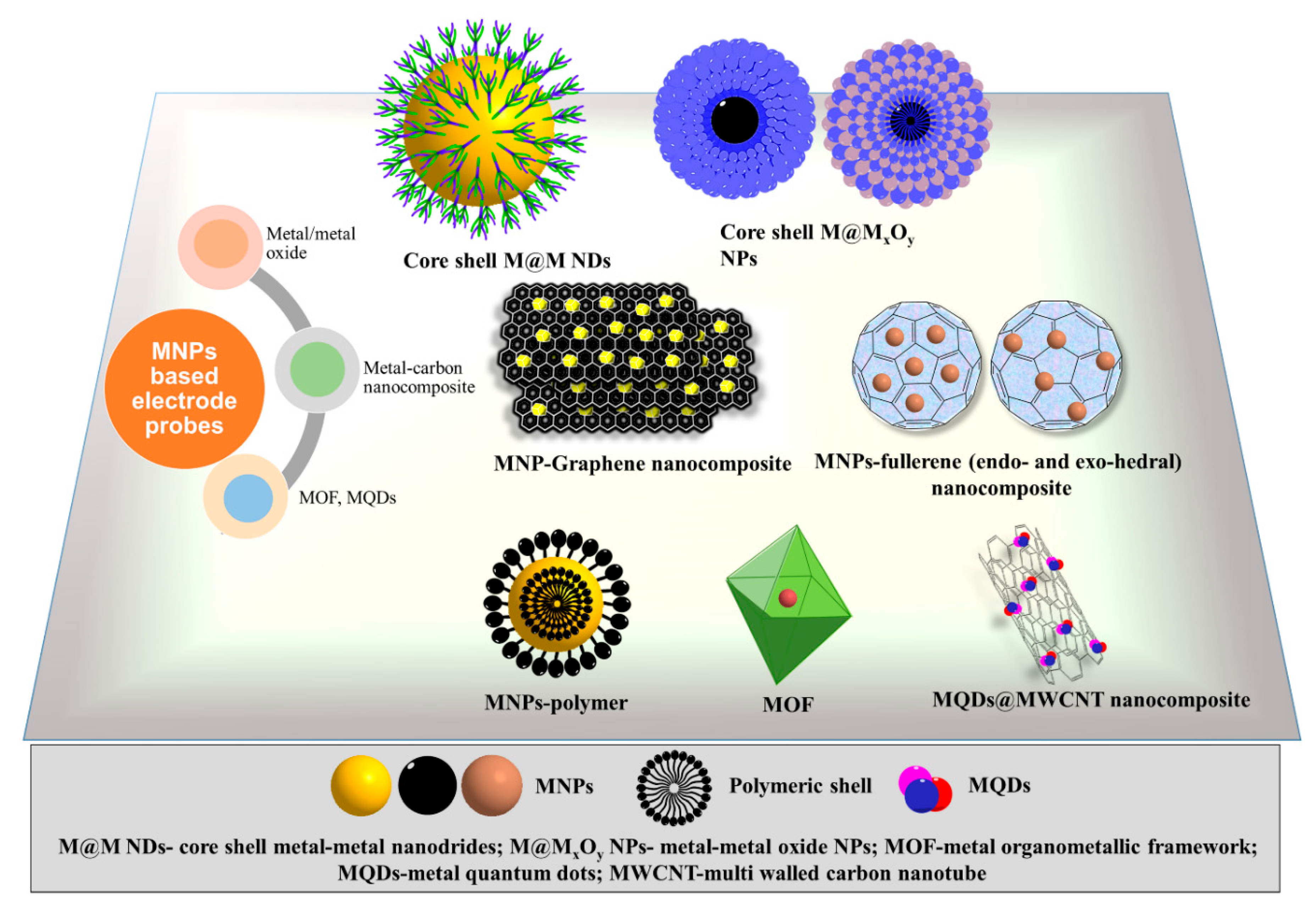
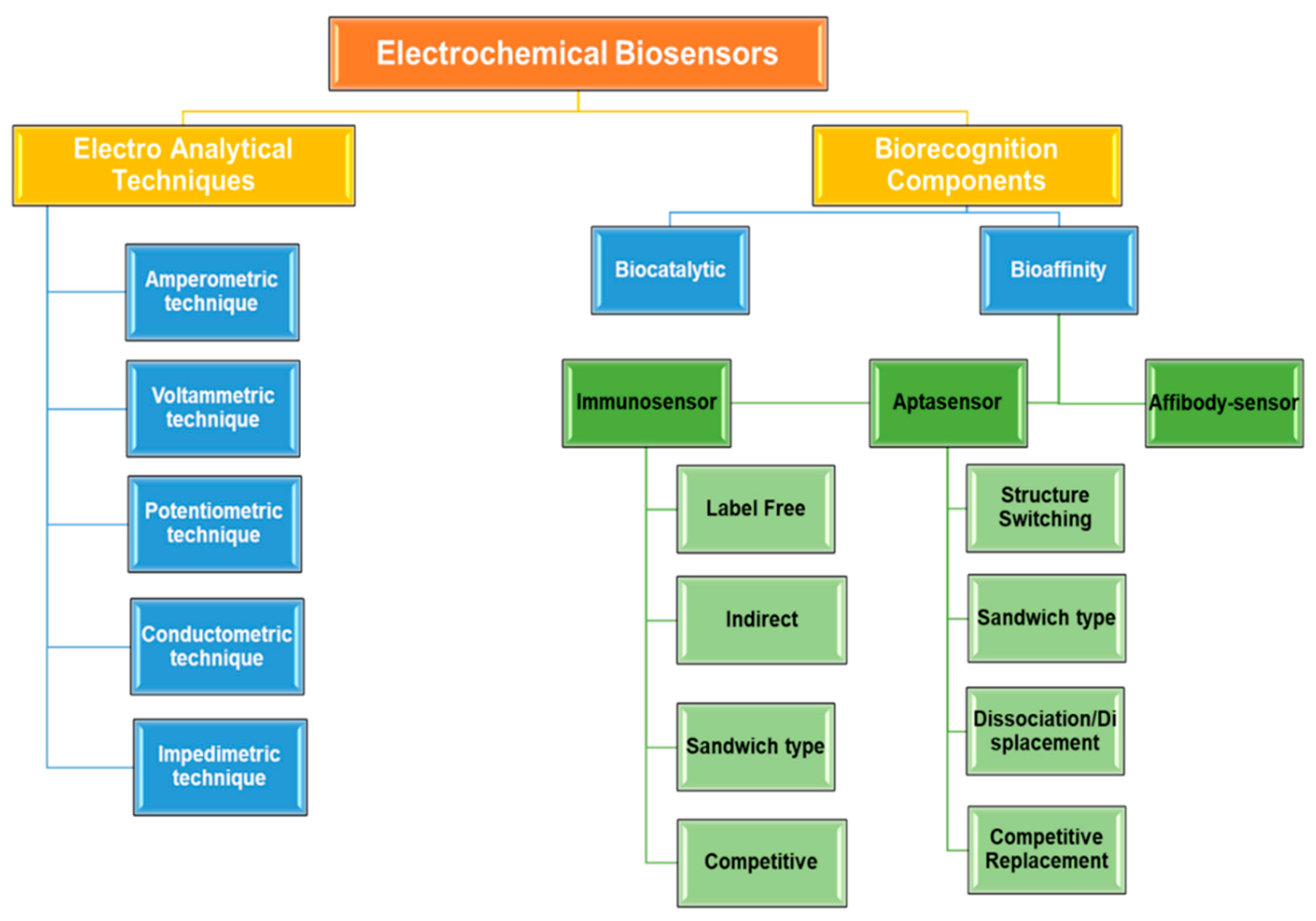
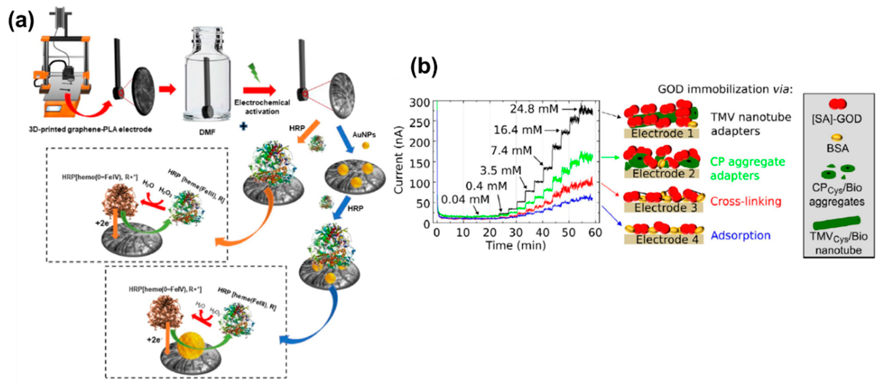
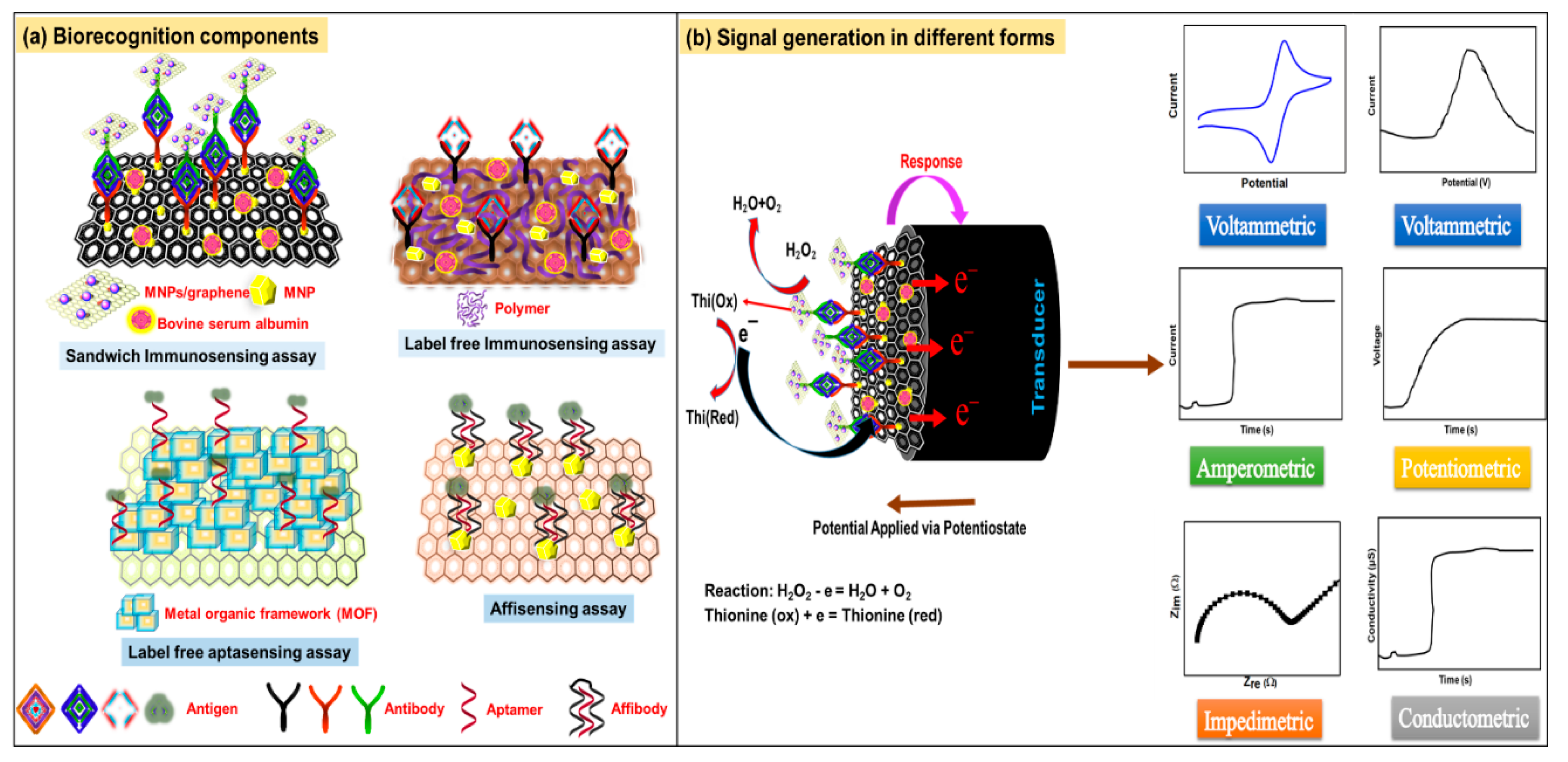
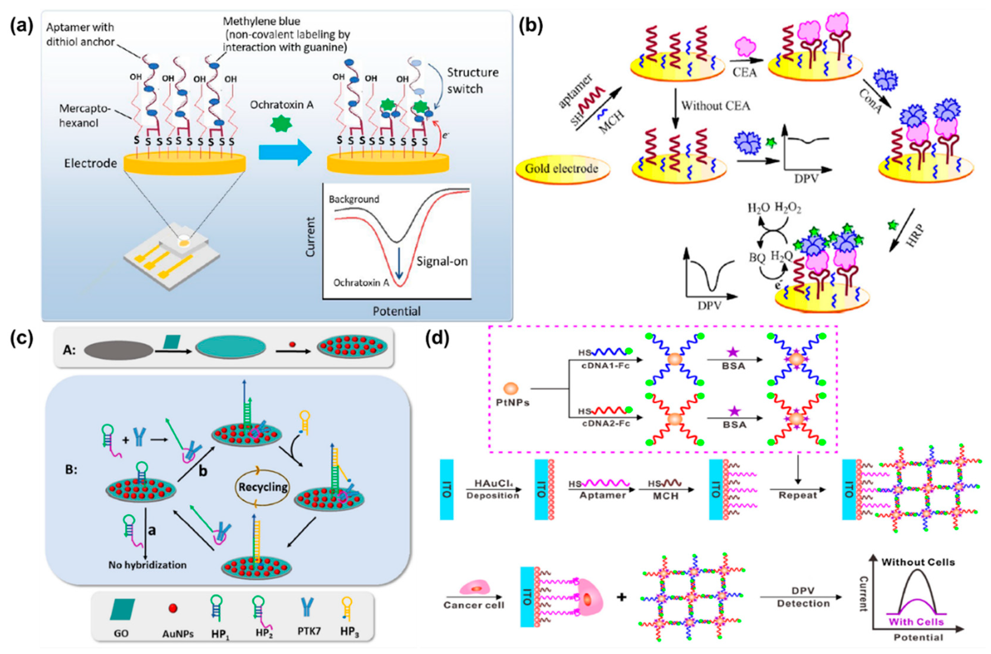
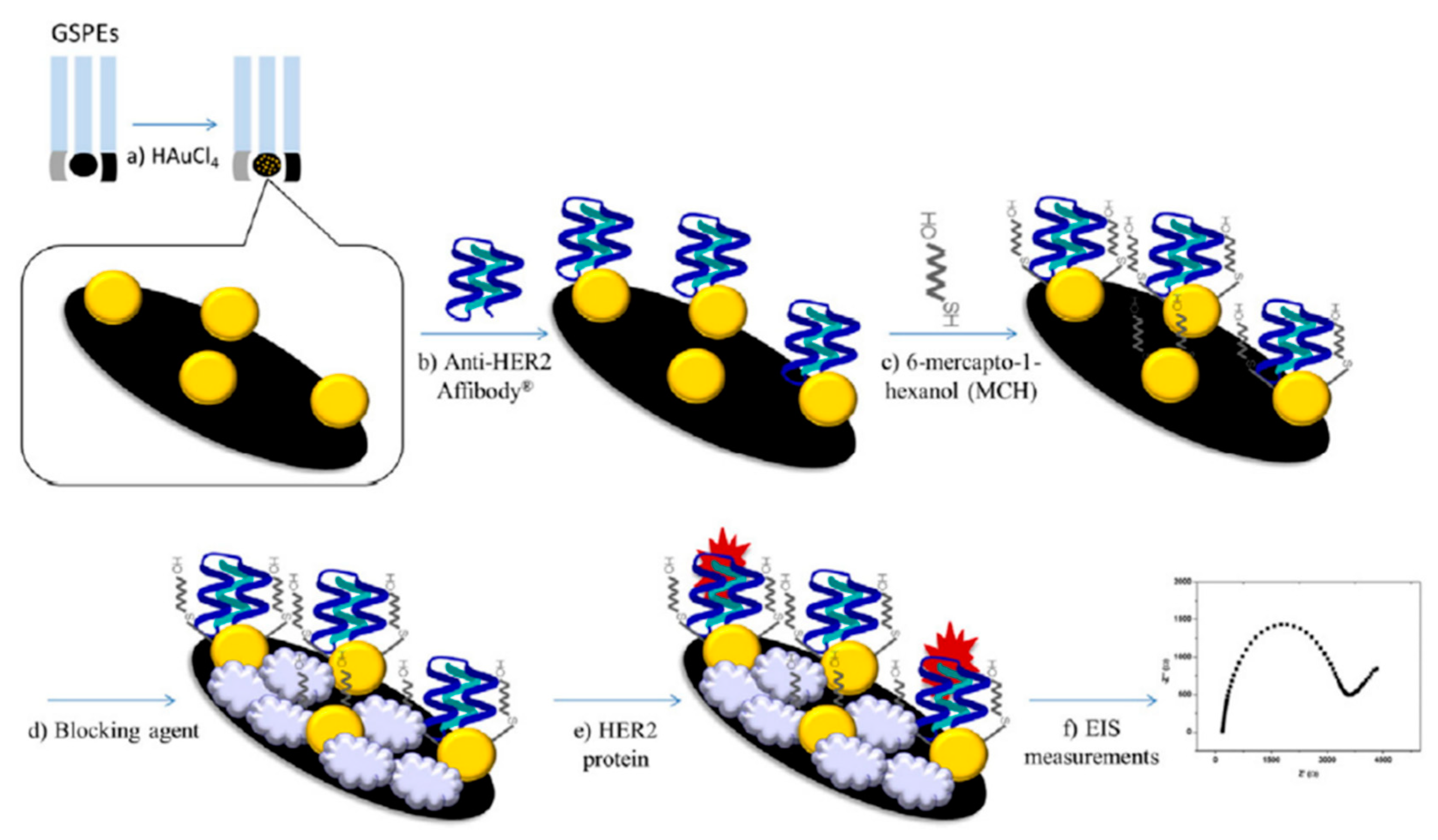
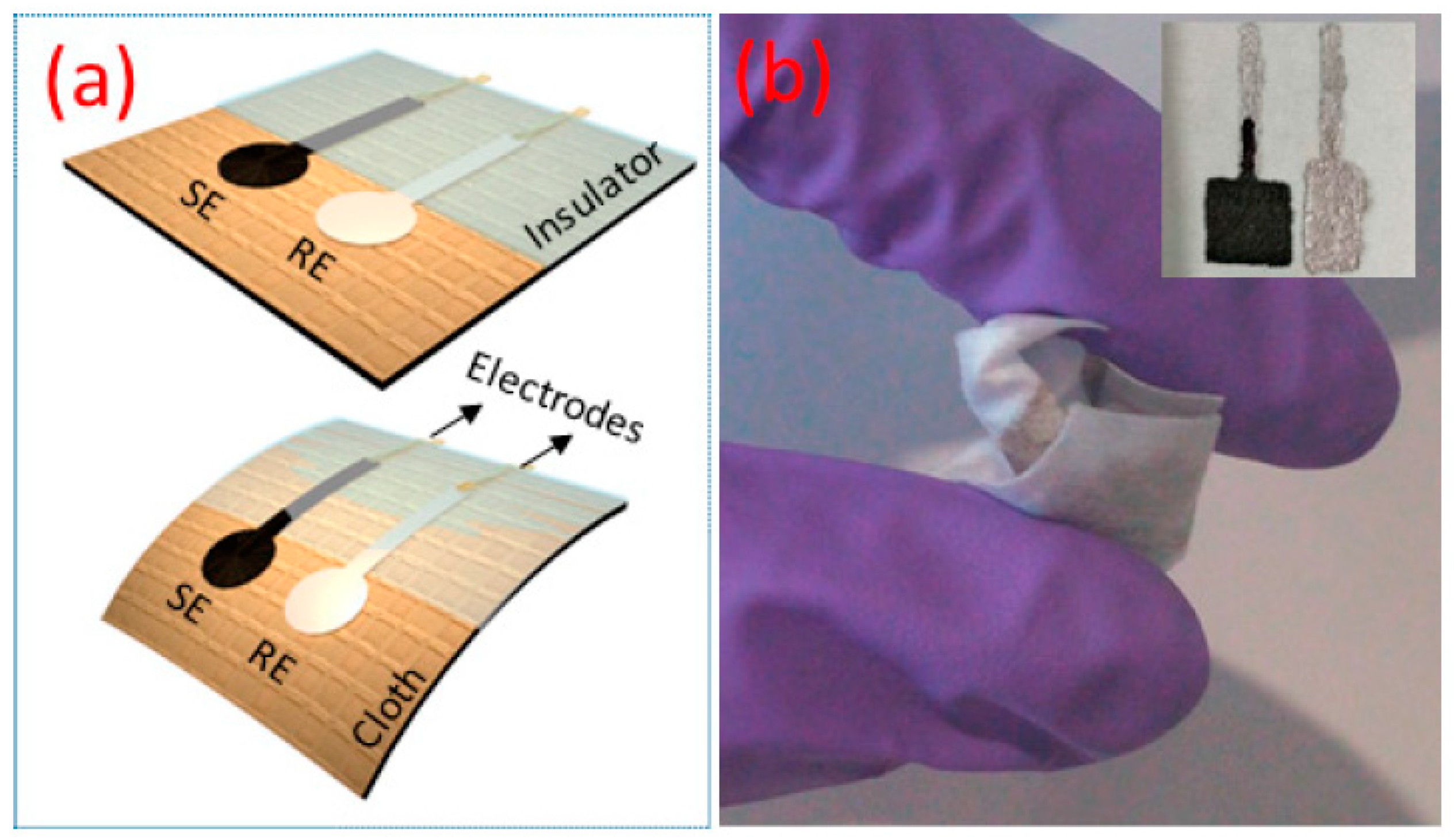
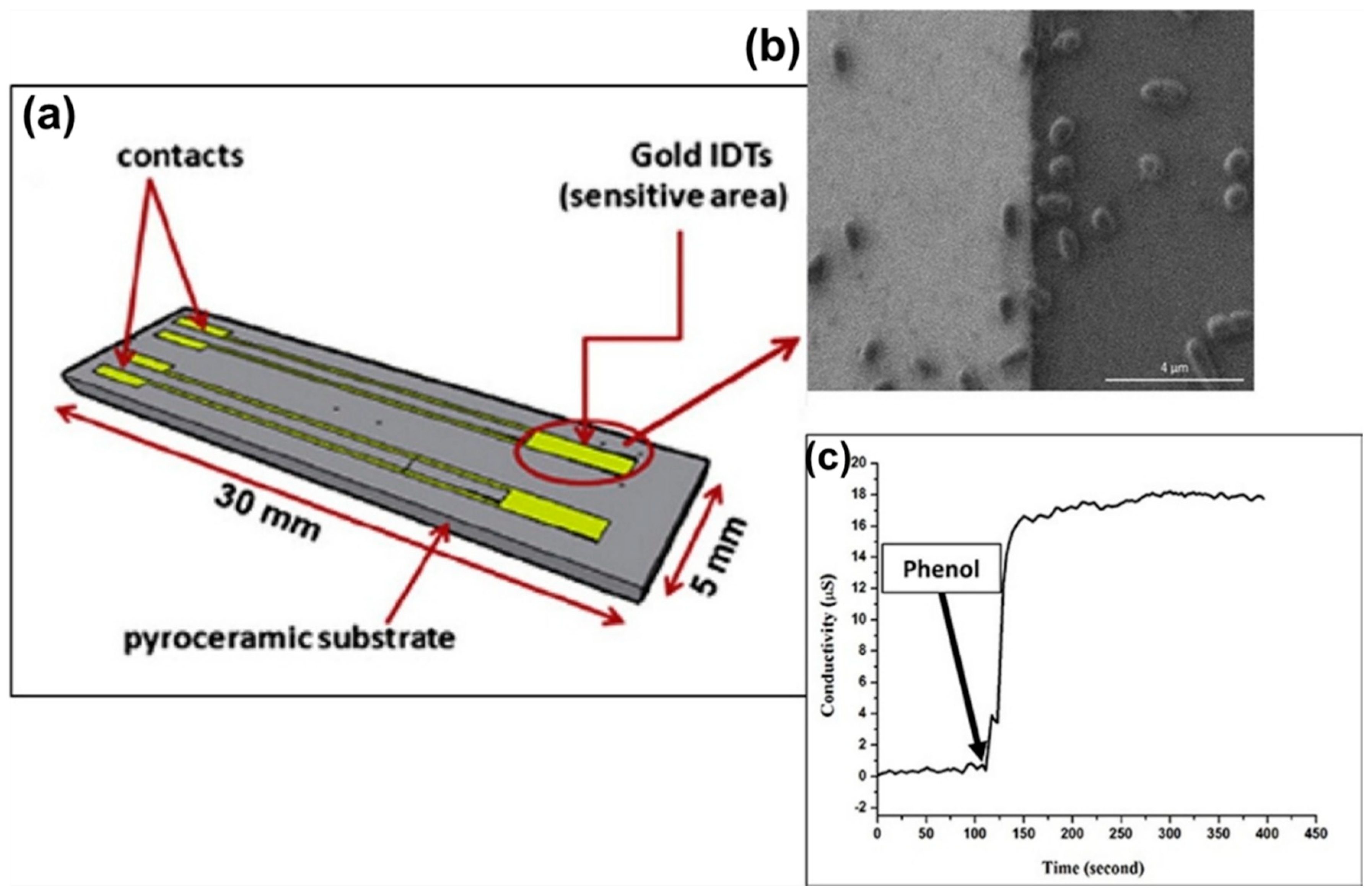
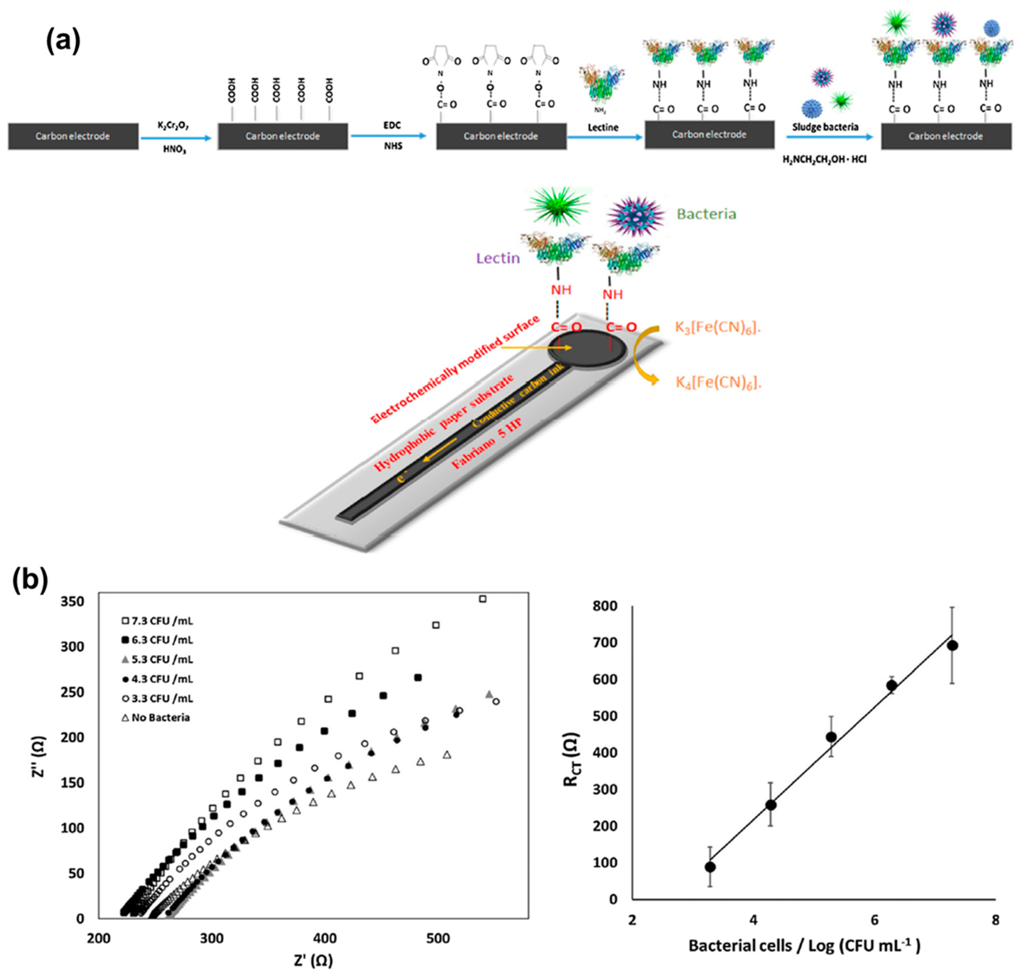
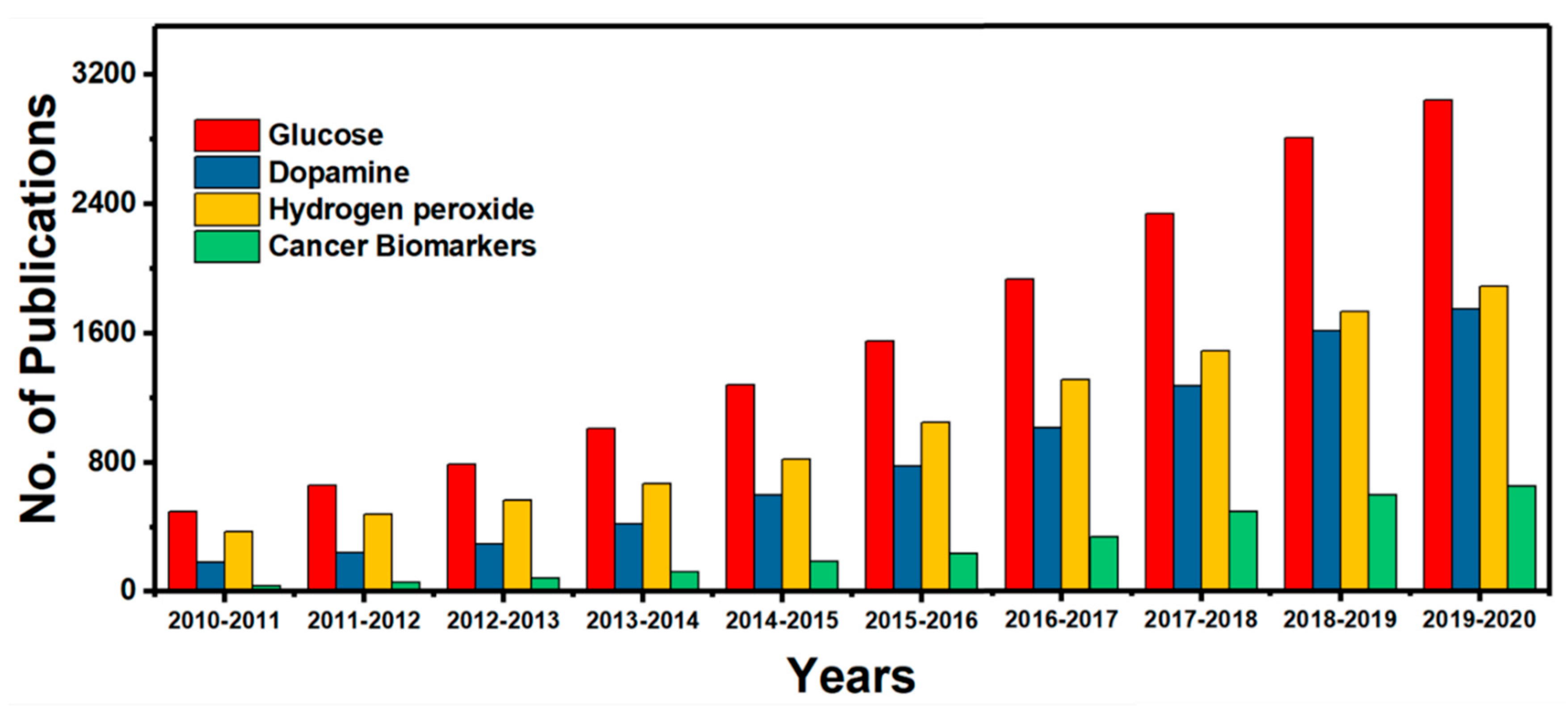
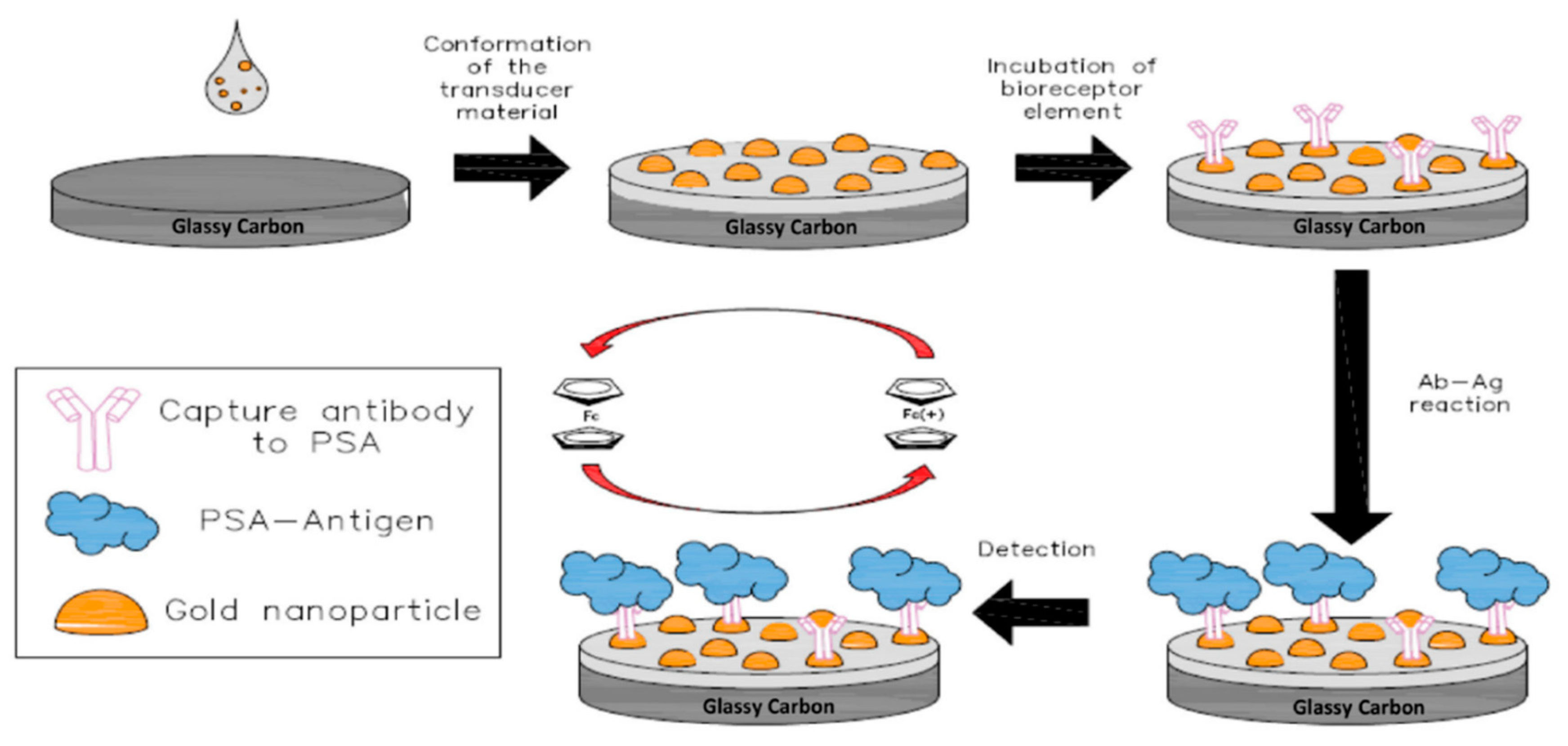
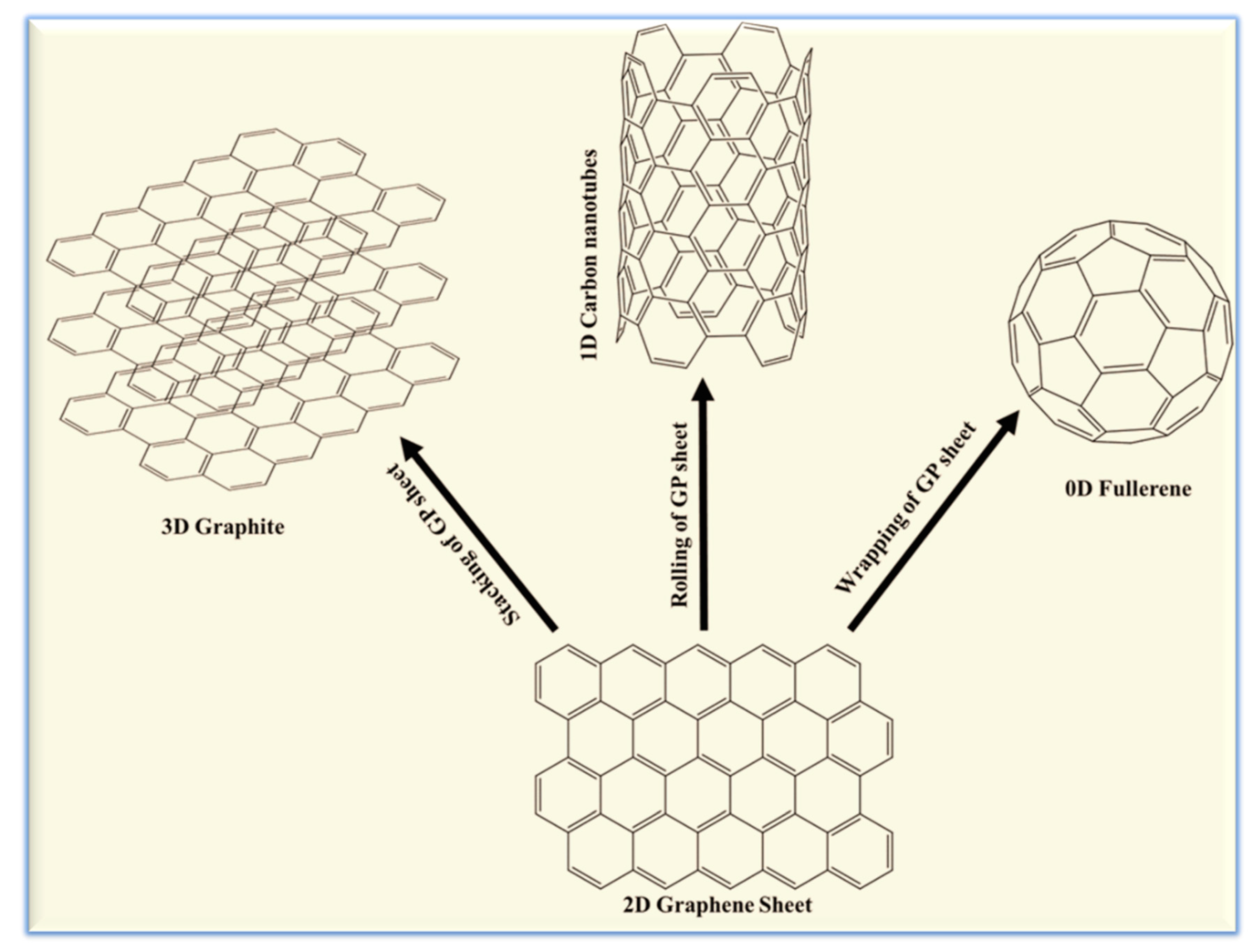
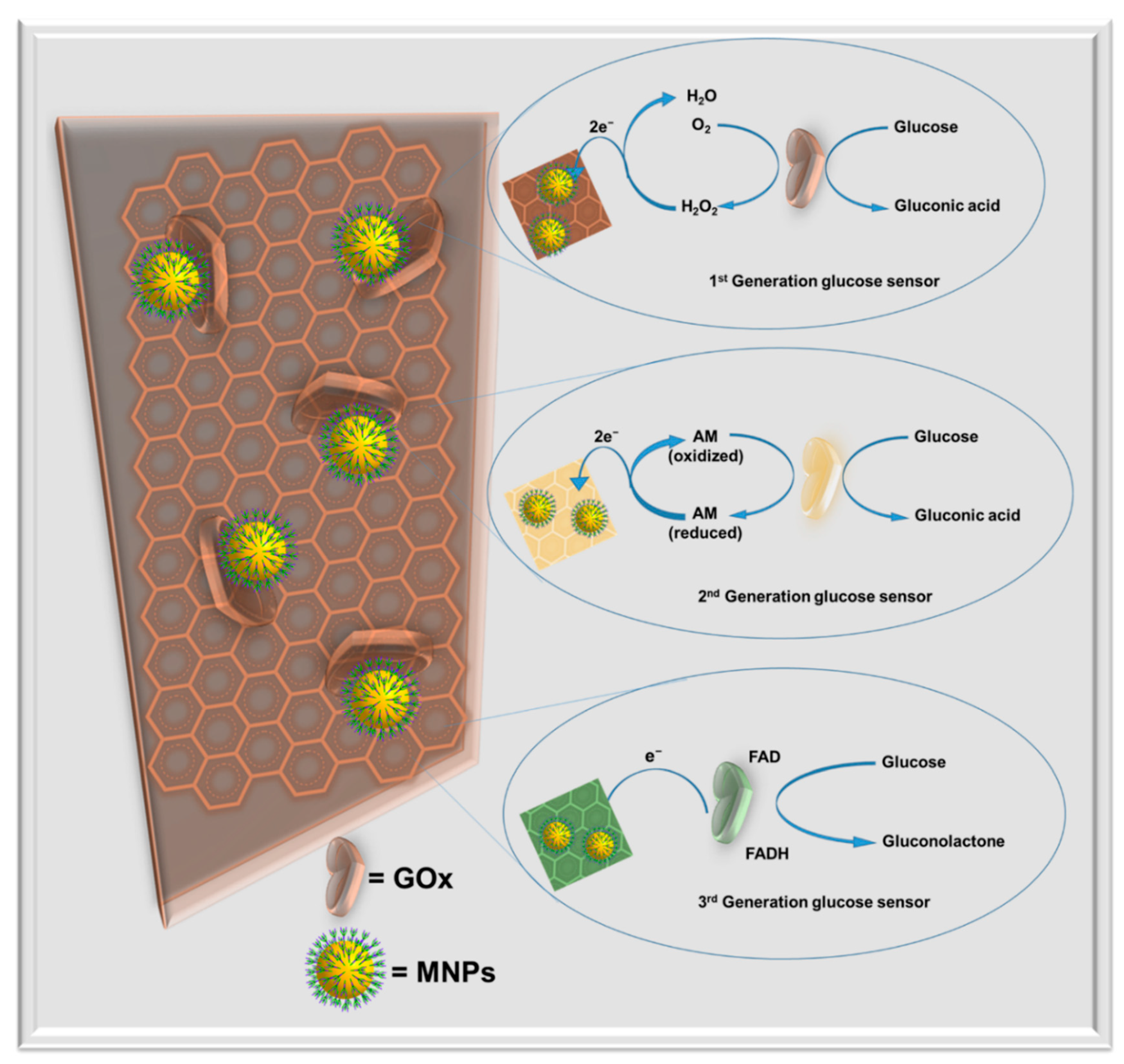
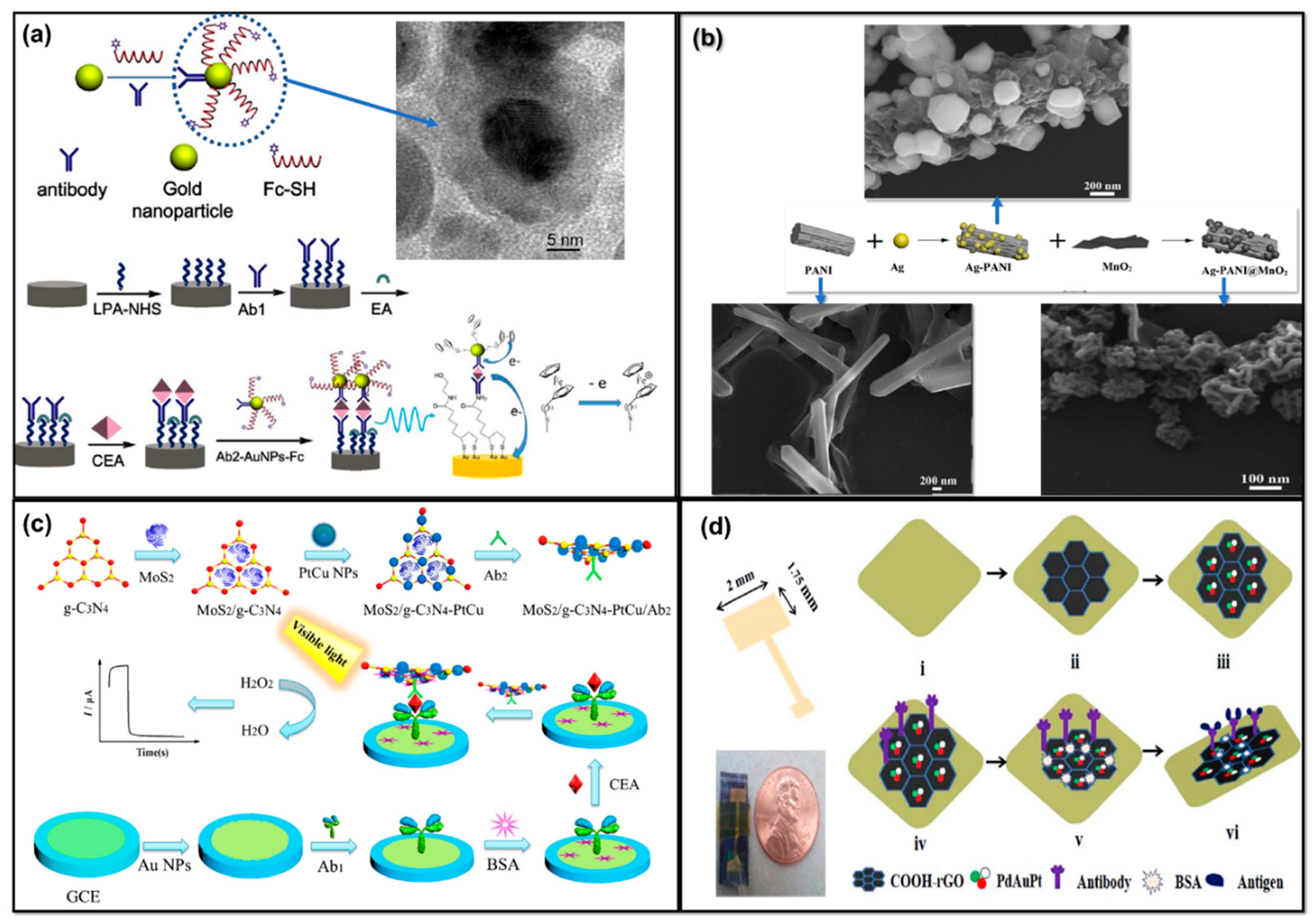
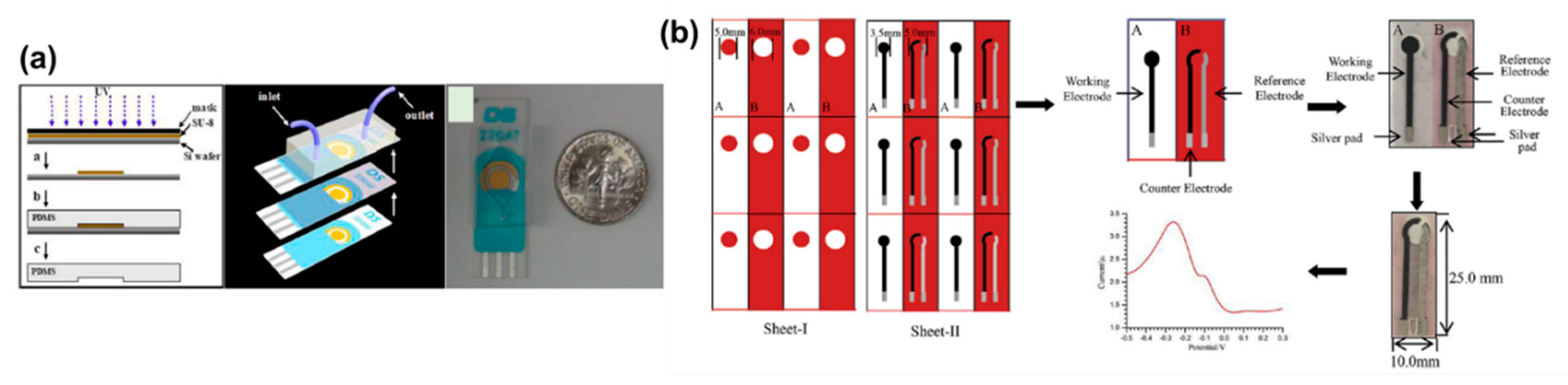
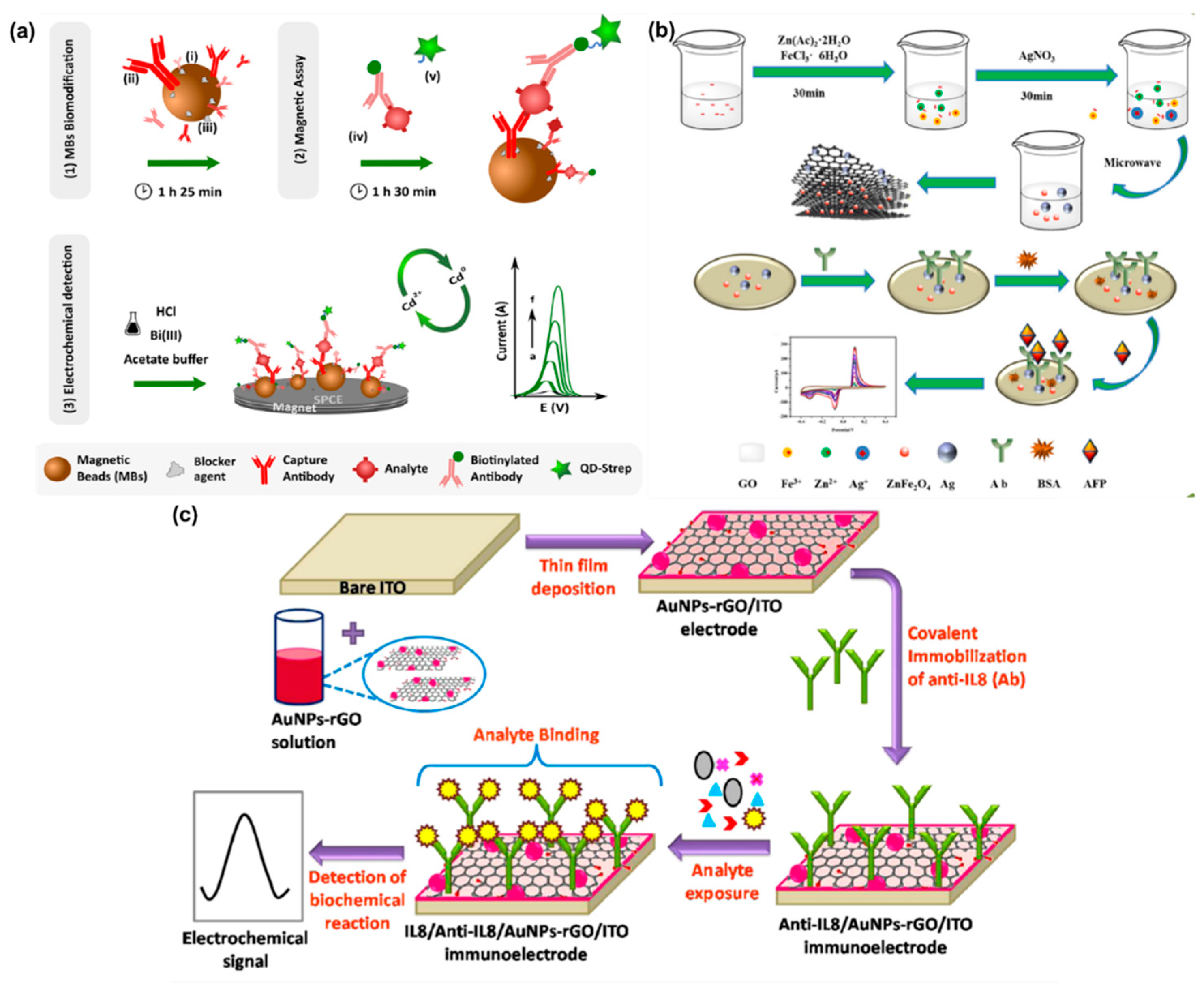
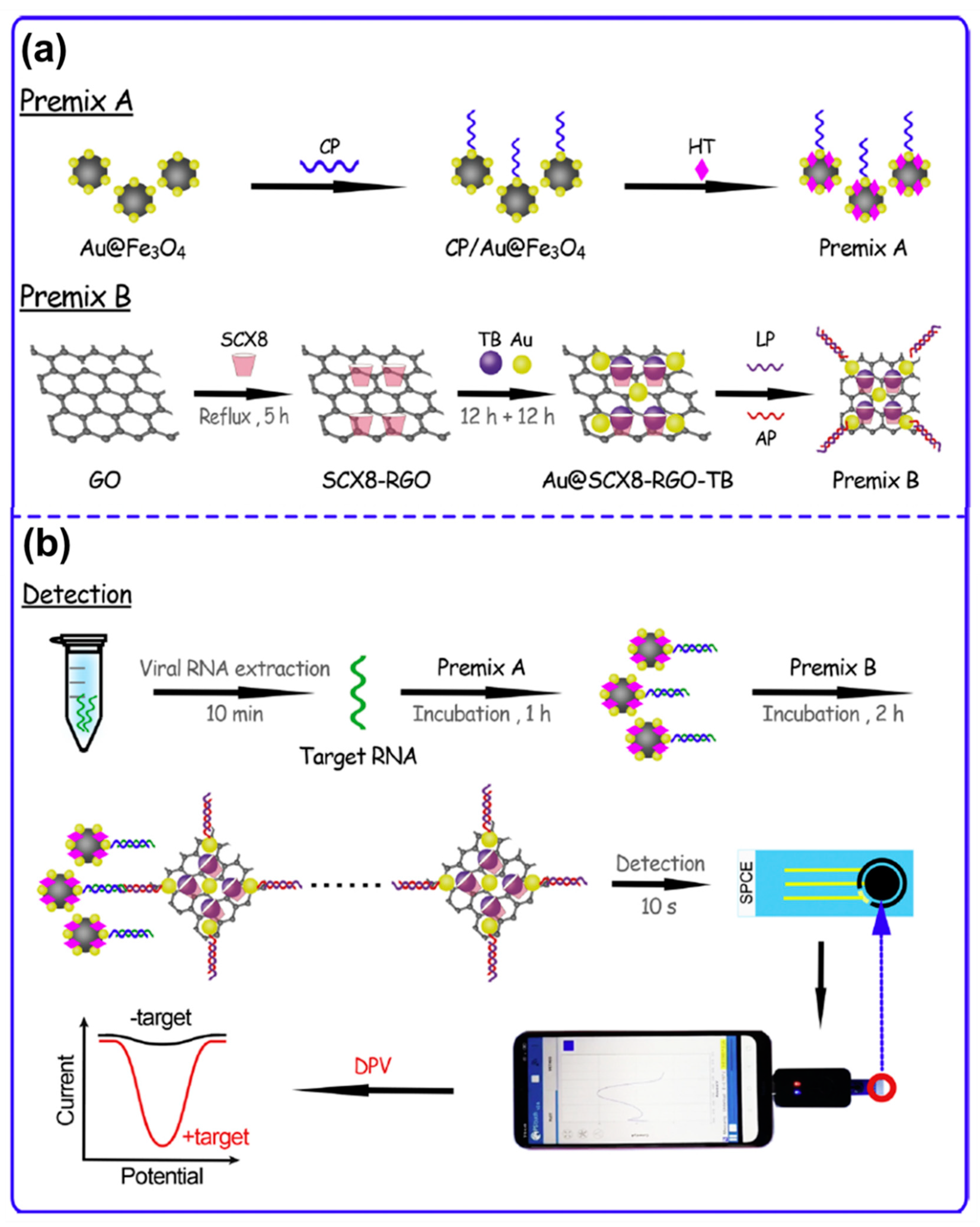
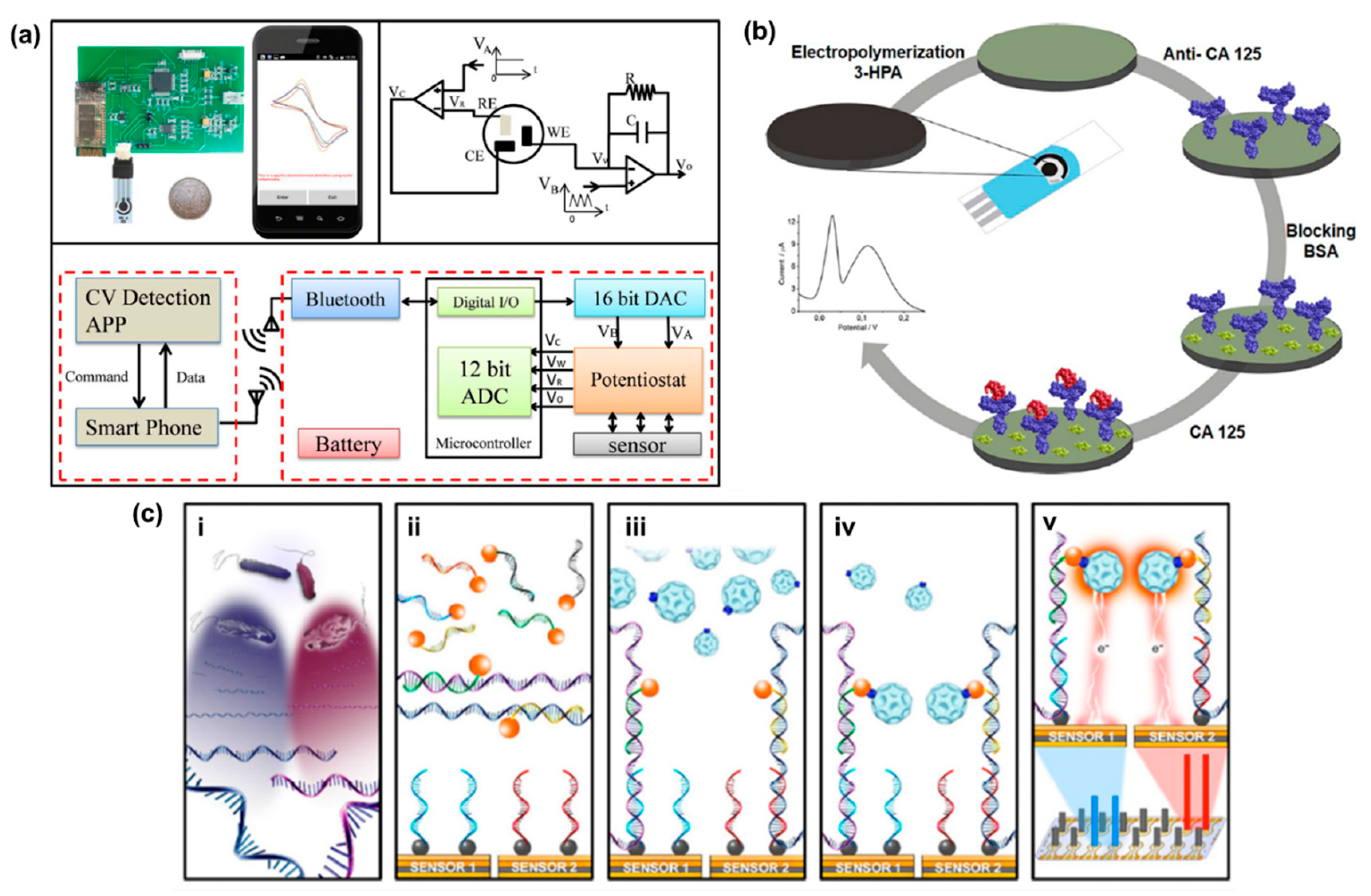
| Transducer | Biorecognition Component | MNP | MNP Size | MNP Shape | Biointeraction and EAT | Analyte | References |
|---|---|---|---|---|---|---|---|
| Ab-N,S-GQDs@AuNP–PANI | anti-HEV antibody | AuNP | 6–14 nm | spherical | Bioaffinity immunosensor, pulse impedance | HEV | [4] |
| AuNP/SWCNTs/PDA/gold electrode | probe DNA | AuNP | 15 nm | oval | Bioaffinity aptasensor, LSV | target DNA | [23] |
| HRP@PGA-C/AgNP | HRP | AgNP | 5–8 nm | spherical | Enzyme-based Biocatalytic, amperometry | H2O2 | [26] |
| PtNPs/MWCNT/PEDOT | glutamate oxidase | PtNP | ~12–20 nm | spherical | Enzyme-based Biocatalytic, amperometry | Glutamate | [27] |
| PtNP/GR/SPCE | ortho-phenylenediamine | PtNP | 400 nm | urchin | Non-enzymatic Biocatalytic, CV | Cotinine | [28] |
| AuNPs–CS/GR/CPE | single-stranded DNA Aptamer | AuNP | 10–20 nm | spherical | Bioaffinity aptasensor, DPV | Activated protein C | [29] |
| Au/AuNP–avidin-HRP | avidin-HRP | AuNP | 40 nm | nanowall | Enzyme-based Biocatalytic, amperometry | DNA Methylation | [39] |
| MWCNT–AuNPs/GCE | E-cadherin antibody–QD | AuNP | 5 nm | spherical | Bioaffinity immunosensor, DPV | Epithelial-mesenchymal transition | [41] |
| Cu–nanoflower@AuNPs–GO NFs coated Au chip | GOx–HRP–Cu nanoflower | AuNP | 20 nm | spherical | Enzyme-based Biocatalytic, amperometry | Glucose | [44] |
| PB–CS–AuNPs/SPCE | HRP-labeled histamine antibody | AuNP | ~50 nm | spherical | Bioaffinity immunosensor, amperometry | HA | [105] |
| DMF–EC/AuNPs/HRP | HRP | AuNP | ~20–30 nm | spherical | Enzyme-based Biocatalytic, amperometry | H2O2 | [62] |
| BSA/anti-A(1–42)/AuNPs/MPA/Au | monoclonal antibody mAb DE2B4 | AuNP | ~30 nm | spherical | Bioaffinity immunosensor, SWV | amyloid beta 1–42 | [70] |
| ds-ATPA/TBA on AuNPs–MoS2/GCE | ds-ATPA and TBA | AuNP | 10 nm | spherical | Bioaffinity aptasensor, SWV | ATP and Thrombin | [85] |
| AgNPs@GQDs/CS/GCE | GOx | AgNP | 40 nm | spherical | Enzyme-based Biocatalytic, CV and amperometry | Glucose | [155] |
| GOx/PtNP@SnS2/Nafion/GCE | GOx | PtNP | ~20–40 nm | spherical | Enzyme-based Biocatalytic, amperometry | Glucose | [168] |
| XO/AuNP/PtNP/MWCNT/GCPE | Xanthine oxidase | AuNP PtNP | 50 nm 5 nm | Spherical spherical | Enzyme-based Biocatalytic, CV | xanthine | [169] |
| AgNPs–Aβ/PrP95–110/GE | peptide | AgNP | ~15 nm | spherical | Bioaffinity aptasensor, LSV | beta-amyloid | [170] |
| Transducer | MNP | Biorecognition Component | Analyte | Linear Range | LOD | References |
|---|---|---|---|---|---|---|
| HRP@PGA-C/AgNP | AgNPs | HRP | H2O2 | 1–3000 µM | 0.35 µM | [26] |
| Cu–nanoflower@AuNPs–GO NFs coated Au chip | AuNPs | GOx–HRP–Cu nanoflower | Glucose | 0.001–0.1 mM | 0.018 µM | [44] |
| DMF-EC/AuNPs/HRP | AuNPs | HRP | H2O2 | 25–100 µM | 9.1 µM | [62] |
| AgNPs@GQDs/CS/GCE | AgNPs | GOx | Glucose | 0.1–10 mM | 0.01 mM | [155] |
| GOx/PtNP@SnS2/Nafion/GCE | PtNPs | GOx | Glucose | 0.1–1 mM 1–12 mM | 2.5 µM | [168] |
| GOx–NiNP/Ni/Au | NiNPs | GOx | Glucose | 1–12 nM | 0.42 mM | [179] |
| Ag-doped PANInanocomposites/GCE | AgNPs | AgNP | DA H2O2 | 10–90 µM 10–90 µM | 0.12 µM 0.03 µM | [183] |
| Ag/MoS2/ITO | AgNPs | Ag/MoS2 | DA | 0.2–50 µM | 0.2 µM | [186] |
| GOx/PtNP/acetic acid-treated LIG | PtNPs | GOx | glucose | 0.3 µM–2.1 mM | 0.3 µM | [187] |
| GOx/PVA-Fe3O4/Sn | Fe3O4 | GOx | glucose | 1–30 mM | 0.5 mM | [188] |
| UOx/EDC:NHS/CZTS/ITO | Cu2ZnSnS4 | UOx | UA | 50–700 μM | 1.3 µM | [184] |
| UOx/Au-rGO/ITO | AuNPs | UOx | UA | 50–800 μM | 7.32 µM | [185] |
| AuNFs/Fe3O4@ZIF-8-MoS2 | AuNPs | Fe3O4@ZIF-8 | H2O2 | 5 µM–120 mM | 0.9 µM | [45] |
| GCE/HUA/HNT/FAD | HNT | HUA and FAD | H2O2 | 1–250 μM | 0.49 µM | [189] |
| Nanostructure Biorecognition Molecules Modified Transducer (Base Electrode *) | Type of ECBs | EAT | Linear Range (ng/mL) | LOD (ng/mL) | Reference |
|---|---|---|---|---|---|
| Carcinoembryonic antigen (CEA): Colorectal, pancreatic, breast, and liver cancers | |||||
| Au*/COOH–rGO/ PdAuPt/antiCEA | Immunosensor | DPV | 0.012–85 | 0.008 | [195] |
| AuNPs/TiO2–GR/HRP–Ab2 and GCE*/AuNPs/Ab1/CEA | HRP-labeled Sandwich immunosensor | DPV | 0.005–200 | 3.33 × 10⁻⁶ | [196] |
| Fc–AuNPs–Ab2 and Au*/LPA–NHS/Ab1/CEA | Fc-labeled Sandwich immunosensor | SWV | 0.05–20 | 0.01 | [197] |
| Ag@CeO2–Au–Ab2 and GCE*/AuNPs/Ab1/CEA | Sandwich immunosensor | CV, EIS | 0.0001–5 | 32 × 10−6 | [198] |
| MoS2/g-C3N4/PtCu/Ab2 and GCE*/AuNPs/Ab1/CEA | Sandwich immunosensor | i-t | 0.0001–80 | 3 × 10⁻⁵ | [199] |
| Fe3O4@AuNPs–DNA(S1)–S2–S3–CEA-Exoll/Hemin | Magnetic aptasensor | DPV | 0.1–200 | 0.0004 | [200] |
| Ag–PANI@MnO2/Ab2 and GCE*/AuNPs/Ab1/BSA/CEA | Sandwich immunosensor | DPV | 0.0005–80 | 0.00017 | [201] |
| Cu–MOFs–TB/PDA/Ab2 and GCE*/MWCNT/CS/Ab1/CEA | TB-labeled sandwich immunosensor | DPV | 2 × 10⁻⁵–200 | 3 × 10⁻⁶ | [202] |
| Mag–SPCE*/AuNP–MnO2/ Fe3O4@Au/antiCEA | Immunosensor | LSV EIS | 0.001–100 | 0.0001 (LSV) 0.0003 (EIS) | [203] |
| GCE*/PDA–rGO/Ag–Au/antiCEA | Immunosensor | CV | 0.001–80 | 2.86 × 10⁻⁴ | [204] |
| GCE*/HNF/AuNP/cMWCNT/antiCEA | immunosensor | EIS | 0.4–125 | 0.09 | [205] |
| CSH/Ab2/BSA and GCE*/MoS2–Au/Ab1/CEA | Sandwich immunosensor | DPV | 0.0001–80 | 3 × 10⁻⁵ | [206] |
| GCE*/Au@PDA@Fe–MOF/NH2–aptamer/BSA/CEA | Aptasensor | DPV | 1 × 10⁻⁶–1000 | 3.3 × 10⁻⁷ | [207] |
| GCE*/CNT@PAMAM/CdSe NP/Ab2/CEA/Ab1/Fe3O4 | Cation-labeled sandwich immunosensor | SWV | 0.005–50 | 0.0017 | [208] |
| MWCNTs/CoS2@PANI/HRP and GCE*/Au/Ab1/BSA/CEA | HRP-labeled sandwich immunosensor | i-t | 0.001–40 | 0.0003 | [209] |
| GCE*/NCMT@Fe3O4@CuSiO3/ConA/CEA/AuNC-aptamer | Cation-labeled aptasensor | DPV | 0.03–6 | 5.38 × 10⁻³ | [210] |
| CeO2-MoS2/Pb2+/Ab2 and GCE*/Au/Ab1/BSA/CEA | Cation-labeled sandwich immunosensor | SWV | 0.001–80 | 0.0003 | [211] |
| MoS2@Cu2O/Fc/Ab2 and GCE*/Au/Ab1/BSA/CEA | Fc-labeled sandwich immunosensor | SWV | 0.001–80 | 3 × 10⁻⁵ | [212] |
| Au*/Ni-Co-PBA/aptamer/CEA | Aptasensor | EIS | 0.001–5 | 7.4 × 10⁻⁷ | [213] |
| GCE*/MWCNT-SO3H/Rh@Pd ND/Ab1/BSA/CEA | Immunosensor | DPV | 2.5 × 10⁻⁵–100 | 8.3 × 10⁻⁶ | [214] |
| GCE*/rGO-PtAu NP/antiCEA | Immunosensor | SWV | 1 × 10⁻⁵–100 | 7 × 10⁻⁶ | [215] |
| Ag-MOF/AuNP/Ab2 and GCE*/MWCNT/Ab1/BSA/CEA | Ag(I)-labeled sandwich immunosensor | DPV | 0.05–120 | 8 × 10⁻⁶ | [216] |
| GCE*/GO-AuNP/antiCEA | Immunosensor | SWV | 1–40 | 0.0158 | [217] |
| AgNP@Strp-HRP/Ab2 and GCE*/T–GO/AuNP@Strp/Ab1/CEA | HRP-labeled sandwich immnusensor | DPV | 0.0001–0.005 | 7.5 × 10⁻⁵ | [218] |
| Fc: ferrocene; LPA-NHS: Lipoic acid N-hydroxysuccinimide ester; rGO: reduced GO; HNF: honey nanofibers; BSA: bovine serum; PAMAM: poly(amidoamine); Strp: streptavidin; | |||||
| Prostate specific antigen (PSA): Prostate cancer | |||||
| Ab2–HRP and GCE*/C60/PANI@PdNP/Ab1/BSA/PSA | HRP-labeled sandwich immnusensor | CV | 0.00016–38 | 1.95 × 10⁻⁵ | [219] |
| GCE*/MoS2–GA@AuNP/antiPSA/BSA | Immunosensor | DPV | 1 × 10⁻⁵–50 | 3 × 10⁻⁶ | [220] |
| Cu3(BTC)2/Ab2 and Au*/PG@PDA/Ab1/CAS/PSA | Sandwich immunosensor | i-t | 0.1–10 | 0.025 | [221] |
| d-Ti3C2Tx@AuNP/Ab2 and GCE*/ATP–GO@AuNP/Ab1/BSA/PSA | Sandwich immunosensor | DPV | 0.00001–0.001 | 3 × 10⁻⁶ | [222] |
| SPCE*/AuNP@aptamer/MCH/PSA | Aptasensor | DPV | 0.001–200 | 7.7 × 10⁻⁵ | [223] |
| GCE*/CeO2–MnO2/antiPSA | Immunosensor | SWV | 0.005–50 | 0.005 | [224] |
| SPCE*/GO/antiPSA | Immunosensor | DPV | 0.75–100 | 0.27 | [225] |
| Au*/MPA/f-PSA/BSA andAu*/MPA/t-PSA/BSA | Immunosensor | EIS | 0.00002–200 | 3 × 10⁻⁶ (f-PSA) 4 × 10⁻⁶ (t-PSA) | [226] |
| Ab2-CdNi QDs and GCE*/Fe3O4@TMU-10(MOF)-CS/Ab1/BSA | QD-labeled sandwich immunosensor | DPV | 0.001–100 | 0.00045 | [227] |
| Ab2-HRP and GCE*/RC60/CuNP@HQ/Ab1/BSA | HRP-labeled Sandwich immunosensor | DPV | 0.005–20 | 0.002 | [228] |
| GCE*/GQDs–CS–Naf-IL/MWCNT–GR–IL/PPY-MoS2–il–AuPt NP/aptamer/PSA | Aptasensor | SWV | 0.0005–0.35 | 0.00014 | [229] |
| GCE*/MWCNT@AuNP-GQD/Ab1/BSA/PSA | Immunosensor | EIS | 0.001–10 | 0.00048 | [230] |
| CPE*/Gr-Fe3O4 NP/antiPSA | Immunosensor | DPV | 1–150 | 0.38 | [231] |
| Au IDE*/16-MUA+EDC/NHS/antiPSA | Immunosensor | f-EIS and nf-EIS | 0.01–100 and 0.5–1000 | 0.01 and 0.5 | [232] |
| Au*/Peptide/GO@AgNP | ECB | LSV | 0.005–20 | 0.00033 | [233] |
| GCE*/rGO-NH2/AgPtPd-COOH/antiPSA | Immunosensor | DPV | 4 × 10⁻⁶–300 | 4 × 10⁻⁶ | [234] |
| GCE*/AuNP/rGO@AuNP/antiPSA | Immunosensor | SWV EIS | 5.5 × 10⁻⁸–0.25; 1–36 (SWV) and 0.0018–41.15 | 0.06; 0.002 (SWV) and 0.006 (EIS) | [235] |
| GCE*/PANI@AuNP/Pep/aptamerPSA/BSA | Aptasensor | DPV | 0.0001–100 | 8.5 × 10⁻⁵ | [236] |
| Ab2-HRP and CASPAuE-MFD*/Magb-Ab1/BSA/PSA | HRP-labeled Sandwich immunosensor | i-t SWV | 0.001–10 | 0.00084 (i-t) 25.4 fM (SWV) | [237] |
| Ab2/AuNP@cys-AgNP@Cu2+ and GCE*/GS@SnO2/Au@Pt/Ab1/BSA/PSA | Cu2+-labeled sandwich immunosensor | SWV | 0.01–100 | 0.00384 | [238] |
| GCE*/[email protected]/antiPSA and GCE*/fMWCNT@AuNP-50/antiPSA | Immunosensor | CV | 0–4 and 0–6 | 85 and 56 | [141] |
| GCE*/AuNP/Ab1/BSA/PSA/Ab2-S0/DNA concatemer(S1-S2)-AgNP Signal probe–DNA concatemer (S1-S2)–AgNP | Ag-labeled sandwich immunosensor | DPV | 0.0001–75 | 3.3 × 10⁻⁵ | [239] |
| Ab2–HRP and SPE*/CS/AuNP/Ab1/BSA/PSA | HRP-labeled Sandwich immunosensor | SWV | 1–18 | 0.001 | [240] |
| SPCE*/rGO@ thionine–AuNP/DNA aptamer/ | Aptasensor | DPV | 0.05–200 | 0.01 | [241] |
| PtNP-Ab2/BSA–CuNP and GCE*/AuNP/Ab1/BSA/PSA | CuNP-labeled sandwich immunosensor | SWV | 0.0005–100 | 14.57 × 10⁻⁵ | [242] |
| Au@Ag-Cu2O/Ab2 and GCE*/Au@N–GQDs/Ab1/BSA/PSA | Sandwich immunosensor | i-t | 1 × 10⁻⁵–100 | 3 × 10⁻⁶ | [243] |
| CAS: casein; MCH: 6-mercapto-1-hexanol; QD: quantum dot; MPA: 3-mercaptopropionic acid; 16-MUA: 16-[Mercaptoundecanoic acid; EDC: N-(3, Dimethylaminopropyl)-N-ethyl-carbodiimidehydrochloride; NHS: Lipoic acid N-hydroxysuccinimide ester; CASPE-MFD: commercially available screen-printed electrode-based microfluidic devices; AE: gold electrode; | |||||
| Cancer Antigen 125 (CA125): Ovarian cancer, breast cancer, lymphoma | |||||
| Ab2–Suc–Chi@MNPs–TB and GCE*/PAMAM/AuNP–3DrGO–MWCNT/Ab1/Glu/CA125 | TB-labeled sandwich immunosensor | SWV | 0.0005–10 and 10–75 U/mL | 0.006 mU/mL | [244] |
| MB–mAb–HRP–CA125 and Au*/Aptamer | HRP-labeled sandwich aptasensor | CV | 2–100 U/mL. | 0.08 U/mL | [245] |
| Ag–PPy–pAb2 and ITO*/MB–mAb1 | Sandwich immunosensor | LSV | 0.001– 300 U/mL | 7.6 mU/mL | [246] |
| Ab2–GPTMS–SiNPs and Au*/f-GNS/Ab1/CA125 | Sandwich immunosensor | DPV | 1 × 10⁻⁹–1 × 10⁻¹⁵ | 1 × 10⁻¹⁵ | [247] |
| TB or Fc–Probe@Au–TiO2@Ab2 and Ta*/BDD/VBG–Au/Ab1–CA125 or CEA | TB/Fc-labeled sandwich immunosensor | DPV | CA125: 0.5–100 mU/mL CEA: 0.0005-0.1 | CA125: 0.09 mU/mL CEA: 0.00015 | [248] |
| SPCE*/rGO/thionine /AuNPs/antiCA125/BSA | POCT immunosensor | DPV | 0.1–200 U/mL | 0.01 U/mL | [249] |
| ITO*/AgNPs–PAN–oxime NFs/aptamer/cDNA–MB/CA125 | MB-labeled aptasensor | DPV | 0.01–350 U/mL | 0.0042 U/mL | [92] |
| GCE*/AgNPs–GQD/antiCA125/BSA | Immunosensor | DPV SWV | 0.01–400 U/mL | 0.01 U/mL | [250] |
| Hollow MB–PDA–Ab2 and GCE*/Au–rGO/Ab1/BSA/CA125 | MB-labeled sandwich immunosensor | DPV | 0.0001–100 U/mL | 336 nU/mL | [251] |
| GCE*/ATA–CNT–MSA; CdS–Ab2/AFP/Ab1; ZnS–Ab2/CEA/Ab1; HgS–Ab2/CA19-9/Ab1; PbS–Ab2/CA125/Ab1 | CdS, ZnS, HgS and PbS-labeled ratiometric multiplex sandwich immunosensor | DPASV | AFP and CEA: 0.0004–10; CA19-9 and CA125: 0.004–100 U/mL | AFP: 0.00011; CEA: 0.0023; CA19-9: 0.68 mU/mL; CA125: 1.4 mU/mL | [252] |
| SPCE*–AuNP/antiCA125/BSA and SPCE*–PtNP/antiCA125/BSA | Immunosensor | EIS | 450–2916 | AuNP: 419; PtNP: 386 | [253] |
| GCE*/Ag–DPA–GQDs/CysA–AuNP/antiCA125/BSA/CA125 | Immunosensor | DPV | 0.001–400 U/mL | 0.001 U/mL | [254] |
| SPE*/Au–AgNPs/antiCA125/BSA (ISA) and SPE*/Au–AgNPs/CysA/antiCA125/BSA (ISB) | Immunosensor | EIS | ISA: 1–500 IU/mL; ISB: 1–1000 IU/mL | 1.03 IU/mL | [255] |
| GCE/FA@H–PANI@CS–HCl/Ab–Ag@Co3O4/BSA/CA125 | FA-labeled immunosensor | DPV | 0.001–25 | 0.00025 | [256] |
| Ab2–AuNPs–LOx and GCE*/GO/MWCNT/AuNPs–CS/Ab1/BSA | Enzymatic immunosensor | i-t | 0.01–0.5 and 0.5–100 U/mL | 0.002 U/mL | [257] |
| AuNP–Ab2–Cd2+ and ITO*/GNR/Ab1/CA125 | Cd2+-labeled sandwich immunosensor | DPV | 20–100 U/mL | 3.4 U/ mL | [258] |
| GCE*/p(CTAB-CS)-AuNP/antiCA125/BSA | Immunosensor | DPV | 0.001–400 U/mL | 0.001 U/mL | [259] |
| GCE*/PDA/ERGO/CysA–AuNPs/antiCA125–HRP/BSA | HRP-labeled immunosensor | SWV | 0.1–400 U/mL | 0.1 U/mL | [260] |
| TB: toluidine blue; MB: methylene blue; GNS: gold Nanostructures; GPTMS: glycidyloxypropyl trimethoxysilane; BDD: boron-doped diamond; VBG: vertical boron doped graphene; FA: ferro-cenecarboxylic acid; pCTAB: poly cetyl trimethylammoniumbromide; AE: gold electrode; GNR: gold nanorods; | |||||
| Human epidermal growth factor receptor 2 (HER2): Breast cancer | |||||
| SPCE*/AuNPs–MPA/NSCeO2/NHS–PEG– Maleimide/antiCA125/BSA | Immunosensor | DPV | 0.001–0.5 and 0.5–20 | 0.0349 | [261] |
| CdSe@ZnS QDs–Ab2 and SPCE*/c-MagBs/Ab1/EA/HER2 | Cd2+-labeled sandwich immunosensor | DPASV | 0.50–50 | 0.29 | [262] |
| AE*/MnFePBA@AuNP/Aptamer/HER2 and AE*/MnFePBA@AuNP/Aptamer/MCF7 | Aptasensor | EIS | HER2: 0.001–1; MCF7: 500-5 × 10⁴ cell/mL | 0.000247; 36 cell/mL | [263] |
| SPCE*/Ab1/HER2–ECD/Ab2/CdSe@ZnS QDs | Cd2+-labeled sandwich immunosensor | DPASV | 10–150 | 2.1 | [264] |
| AE*/CDs@ZrHf–MOF/Aptamer/HER2 | Aptasensor | EIS | 0.001–10; 1000–1 × 10⁵ cell/mL | HER2: 19 × 10−6; MCF7: 23 cell/mL | [265] |
| Ab2–CDI–PbS QDs and SPCE*–COOH/EDC–NHS/Ab1 | Pb2+-labeled sandwich immunosensor | SWV | 1–100 | 0.28 | [266] |
| GCE*/ErGO–SWCNT/AuNP/Aptamer/HER2 | Aptasensor | EIS | 0.0001–1 | 5 × 10⁻⁵ | [267] |
| GNR–Pd SS–Aptamer–HRP and AE*/DNA tetrahedron/BSA/HER2 | HRP-labeled sandwich aptasensor | DPV | 10–200 | 0.15 | [268] |
| ITO*/MoO3@rGO/APTES/antiHER2/BSA | Immunosensor | DPV | 0.001–500 | 0.001 | [269] |
| GSPE*/AuNPs/antiHER2 affibody/MCH/HER2 | Affisensor | EIS | 0–4 × 10⁴ | 6000 | [100] |
| Ab2–AuNPs–dC20 AE*/peptide/MCH/HER2 | DNA-labeled sandwich immunosensor | SWV | 0.0001–1 | 0.0005 | [270] |
| Ab2/Hyd@AuNPs–APTMS–Fe3O4 and GCE*/Fe3O4–APTMS/Ab1/BSA/HER2 | Sandwich immunosensor | DPV | 0.0005–50 | 2 × 10⁻⁵ | [271] |
| PEG: polyethylene glycol; MagB: magnetic bead; CDI: carbonyldi-imidazole; MCH: 6-mercapto-1-hexanol; APTES: 3-aminopropyltriethoxysilane; APTMS: 3-aminopropyltrimethoxysilane; AE: gold electrode; | |||||
| α-fetoprotein (AFP): Gastrointestinal tumor and liver cancer | |||||
| GCE*/PGNR/AuNPs/antiAFP/BSA/AFP | Immunosensor | DPV | 5–60 | 1.0 | [272] |
| Thiolated LAPS*/MPTES–AuNPs/Aptamer/AFP | Aptasensor | potentiometric | 100–1 × 10⁵ | 92 | [273] |
| AuNP–DNA2–MB and AE*/DNA1/MCH/Fc–CP/AFP/RecJf | Ratiometric aptasensor | ACV | 1 × 10⁻⁵–100 | 269.4 ag/mL | [274] |
| AE*/AlCu MOF2.5,2.5/Aptamer/AFP | Aptasensor | EIS | 0.001–0.5 | 0.00012 | [275] |
| Pd@PtNPs–Ab2– thionine and AE*/AuNPs/Ab1/BSA/AFP | Thionine-labeled sandwich immunosensor | DPV | 0.0001–100 | 0.000035 | [276] |
| Ab2–AgNP–HRP and GCE*/PANI–AgNP/Ab1/BSA/AFP | HRP-labeled sandwich immunosensor | i-t | 0.01–1 and 1–10 | 0.0047 | [277] |
| GCE*/ZnFe2O4–AgNP@rGO/antiAFP/BSA/AFP | Label-free immunosensor | CV | 0.001–200 | 0.00098 | [278] |
| GCE*/Cu3Pt NPs/antiAFP/BSA/AFP | Immunosensor | DPV | 0.0001–10 | 0.000033 | [279] |
| MO/CNT–AuNP–Ab2 and GCE*/VG–AuPt/Ab1/BSA/AFP | MO-labeled sandwich immunosensor | DPV | 1 × 10⁻⁶–100 | 7 × 10⁻⁷ | [280] |
| MoSe2 NSs–NH2/Au@Pt DNRs–Ab2 and GCE*/GS–NH2/AuNPs/Ab1/BSA/AFP | Sandwich immunosensor | i-t | 1 × 10⁻⁵–200 | 3.3 × 10⁻⁶ | [281] |
| PGNR: porous graphene nanoribbon; MPTES: 3-mercaptopropyltriethoxysilane; AE: gold electrode; BSA: bovine serum; MO: methyl orange; DNR: dendritic nanorods; | |||||
| Interleukin-6 (IL-6): Colorectal cancer | |||||
| MCH/Apt/AuNPs/pATP/pABA/GCE | Sandwich aptasensor | EIS | 0.005–100 | 0.0016 | [282] |
| MCHApt/AuNPs/PPyNPs/SPGE | Structure switching aptasensor | EIS | 0.001–15000 | 0.00033 | [283] |
| MCH: 6-mercapto-1-hexanol; Apt: aptamer; pATP: p-aminothiophenol; pABA: p-aminobenzoic acid; PPY: polypyrrole; | |||||
| Interleukin-8 (IL-8): Oral cancer | |||||
| BSA/Anti-IL 8/β–Ag2MoO4/ITO | Label-free immunosensor | DPV | 1 × 10⁻⁶–40 | 0.09 | [284] |
| DNA-templated CdTe/CdS QDs/MB | Aptasensor | ASV | 1 × 10⁻⁶–0.005 | 3.36 × 10⁻⁶ | [285] |
| Anti-IL8/AuNPs–rGO/ITO | Label-free immunosensor | DPV | 0.5–4 | 0.072 | [286] |
| BSA: bovine serum; MB: methylene blue; ITO: indium tin oxide; | |||||
Publisher’s Note: MDPI stays neutral with regard to jurisdictional claims in published maps and institutional affiliations. |
© 2020 by the authors. Licensee MDPI, Basel, Switzerland. This article is an open access article distributed under the terms and conditions of the Creative Commons Attribution (CC BY) license (http://creativecommons.org/licenses/by/4.0/).
Share and Cite
Islam, T.; Hasan, M.M.; Awal, A.; Nurunnabi, M.; Ahammad, A.J.S. Metal Nanoparticles for Electrochemical Sensing: Progress and Challenges in the Clinical Transition of Point-of-Care Testing. Molecules 2020, 25, 5787. https://doi.org/10.3390/molecules25245787
Islam T, Hasan MM, Awal A, Nurunnabi M, Ahammad AJS. Metal Nanoparticles for Electrochemical Sensing: Progress and Challenges in the Clinical Transition of Point-of-Care Testing. Molecules. 2020; 25(24):5787. https://doi.org/10.3390/molecules25245787
Chicago/Turabian StyleIslam, Tamanna, Md. Mahedi Hasan, Abdul Awal, Md Nurunnabi, and A. J. Saleh Ahammad. 2020. "Metal Nanoparticles for Electrochemical Sensing: Progress and Challenges in the Clinical Transition of Point-of-Care Testing" Molecules 25, no. 24: 5787. https://doi.org/10.3390/molecules25245787